AICDA antibody
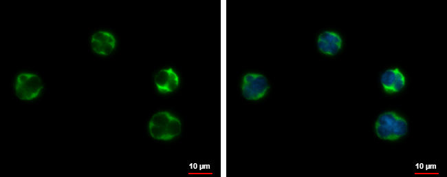
AICDA antibody detects AICDA protein at cytoplasm by immunofluorescent analysis.
Sample: Raji cells were fixed in 4% paraformaldehyde at RT for 15 min.
Green: AICDA protein stained by AICDA antibody (GTX127276) diluted at 1:500.
Blue: Hoechst 33342 staining.
Scale bar = 10 μm.
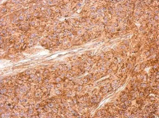
AICDA antibody detects AICDA protein at cytosol on SAS xenograft by immunohistochemical analysis.
Sample: Paraffin-embedded SAS xenograft.
AICDA antibody (GTX127276) dilution: 1:500.
Antigen Retrieval: Trilogy™ (EDTA based, pH 8.0) buffer, 15min
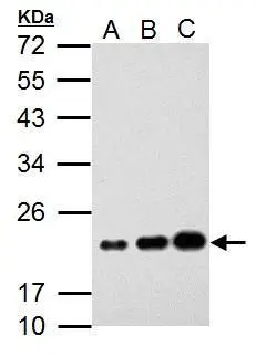
Sample (30 μg of whole cell lysate)
A: K562
B: THP-1
C: HL-60
12% SDS PAGE
GTX127276 diluted at 1:1000
The HRP-conjugated anti-rabbit IgG antibody (GTX213110-01) was used to detect the primary antibody.
-
HostRabbit
-
ClonalityPolyclonal
-
IsotypeIgG
-
ApplicationsWB ICC/IF IHC-P
-
ReactivityHuman