AKT2 antibody


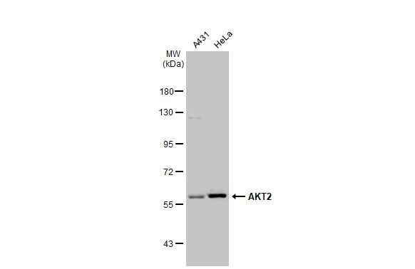
Various whole cell extracts (30 μg) were separated by 7.5% SDS-PAGE, and the membrane was blotted with AKT2 antibody (GTX128458) diluted at 1:1000. The HRP-conjugated anti-rabbit IgG antibody (GTX213110-01) was used to detect the primary antibody.
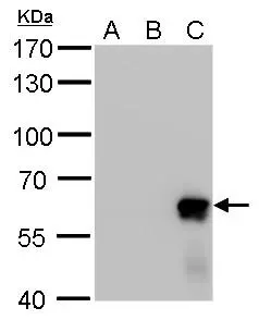
Akt2 antibody detects Akt2 protein by Western blot analysis.
A. 30 μg 293T whole cell lysate/extract
B. 30 μg whole cell lysate/extract of HA-human Akt1-transfected 293T cells
C. 5 μg whole cell lysate/extract of HA-human Akt2-transfected 293T cells
7.5 % SDS-PAGE
Akt2 antibody (GTX128458) dilution: 1:20000
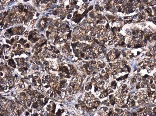
AKT2 antibody detects AKT2 protein at cytoplasm and nucleus in human oral carcinoma by immunohistochemical analysis.
Sample: Paraffin-embedded human oral carcinoma.
AKT2 antibody (GTX128458) diluted at 1:400.
Antigen Retrieval: Citrate buffer, pH 6.0, 15 min
-
HostRabbit
-
ClonalityPolyclonal
-
IsotypeIgG
-
ApplicationsWB IHC-P
-
ReactivityHuman