AKT3 antibody

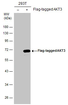
Non-transfected (–) and transfected (+) 293T whole cell extracts (30 μg) were separated by 10% SDS-PAGE, and the membrane was blotted with AKT3 antibody (GTX113312) diluted at 1:1000. The HRP-conjugated anti-rabbit IgG antibody (GTX213110-01) was used to detect the primary antibody.
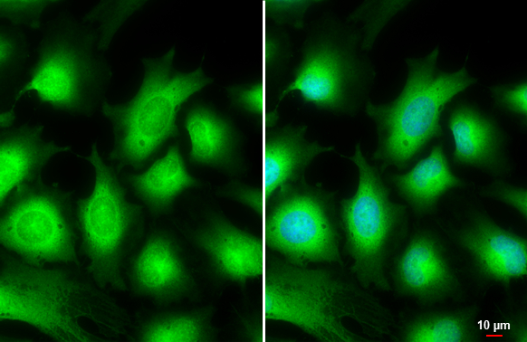
AKT3 antibody detects AKT3 protein at cytoplasm by immunofluorescent analysis.Sample: HeLa cells were fixed in 4% paraformaldehyde at RT for 15 min.Green: AKT3 stained by AKT3 antibody (GTX113312) diluted at 1:500.Blue: Hoechst 33342 staining.Scale bar= 10 μm.
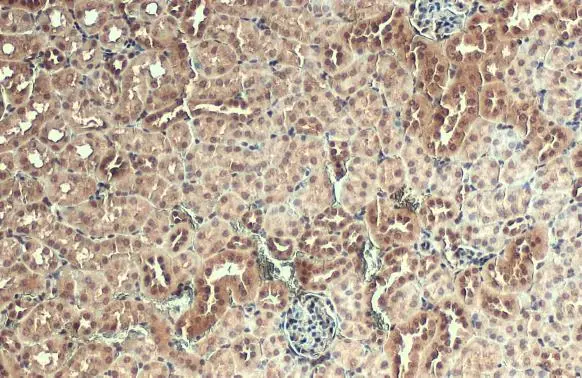
AKT3 antibody detects AKT3 protein at nucleus by immunohistochemical analysis.Sample: Paraffin-embedded mouse kidney.AKT3 stained by AKT3 antibody (GTX113312) diluted at 1:500.Antigen Retrieval: Citrate buffer, pH 6.0, 15 min
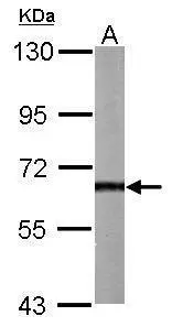
Sample (50 μg of whole cell lysate)
A: Mouse brain
7.5% SDS PAGE
GTX113312 diluted at 1:1000
The HRP-conjugated anti-rabbit IgG antibody (GTX213110-01) was used to detect the primary antibody.
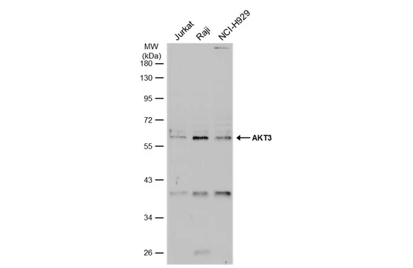
Various whole cell extracts (30 μg) were separated by 10% SDS-PAGE, and the membrane was blotted with AKT3 antibody (GTX113312) diluted at 1:1000. The HRP-conjugated anti-rabbit IgG antibody (GTX213110-01) was used to detect the primary antibody, and the signal was developed with Trident ECL plus-Enhanced.
-
HostRabbit
-
ClonalityPolyclonal
-
IsotypeIgG
-
ApplicationsWB ICC/IF IHC-P
-
ReactivityHuman, Mouse