APE1 antibody

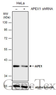
Non-transfected (–) and transfected (+) HeLa whole cell extracts (30 μg) were separated by 10% SDS-PAGE, and the membrane was blotted with APE1 antibody (GTX110558) diluted at 1:1000. The HRP-conjugated anti-rabbit IgG antibody (GTX213110-01) was used to detect the primary antibody.
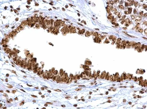
APE1 antibody detects APE1 protein at cytoplasm, nucleus and nucleolus on human ovarian carcinoma by immunohistochemical analysis.
Sample: Paraffin-embedded human ovarian carcinoma.
APE1 antibody (GTX110558) diluted at 1:500.
Antigen Retrieval: Trilogy™ (EDTA based, pH 8.0) buffer, 15min
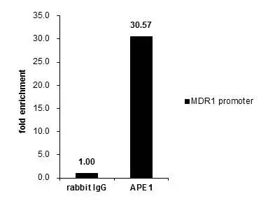
ChIP experiment and primer designs are based on Oncogene. 2011 Jan 27;30(4):482-93.
Cross-linked ChIP was performed with 293T chromatin extract treated with Trichostatin A (0.4 μM for 18 h) and 5 μg of either control rabbit IgG or anti-APE1 antibody. The precipitated DNA was detected by PCR with primer set targeting to MDR1 promoter.
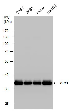
APE1 antibody detects APE1 protein by Western blot analysis. Various whole cell extracts (30 μg) were separated by 10% SDS-PAGE, and the membrane was blotted with APE1 antibody (GTX110558) diluted by 1:1000.
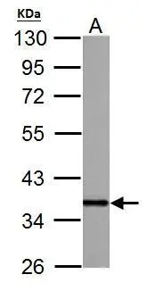
APE1 antibody detects APEX1 protein by western blot analysis.
A. 30 μg PC-12 whole cell lysate/extract
10% SDS-PAGE
APE1 antibody (GTX110558) dilution: 1:10000
The HRP-conjugated anti-rabbit IgG antibody (GTX213110-01) was used to detect the primary antibody.
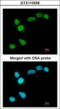
Immunofluorescence analysis of paraformaldehyde-fixed A549, using APE1(GTX110558) antibody at 1:200 dilution.
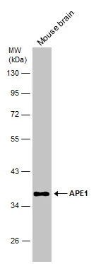
Mouse tissue extract (50 μg) was separated by 10% SDS-PAGE, and the membrane was blotted with APE1 antibody (GTX110558) diluted at 1:1000. The HRP-conjugated anti-rabbit IgG antibody (GTX213110-01) was used to detect the primary antibody.
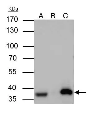
APE1 antibody immunoprecipitates APE1 protein in IP experiments. IP Sample: A431 whole cell lysate/extract A : 30 μg whole cell lysate/extract of APE1 protein expressing A431 cells B : Control with 3 μg of pre-immune rabbit IgG C : Immunoprecipitation of APE1 by 3 μg of APE1 antibody (GTX110558) 10% SDS-PAGE The immunoprecipitated APE1 protein was detected by APE1 antibody (GTX110558) diluted at 1 : 1000. EasyBlot anti-rabbit IgG (HRP) (GTX221666-01) was used as a secondary reagent.
-
HostRabbit
-
ClonalityPolyclonal
-
IsotypeIgG
-
ApplicationsWB ICC/IF IHC-P IP ChIP assay
-
ReactivityHuman, Mouse, Rat