APPLICATION
Application Note
*Optimal dilutions/concentrations should be determined by the researcher.
Application |
Recommended Dilution |
1:500 - 1:2000 |
1:50 - 1:100 |
1:50 - 1:200 |
Not tested in other applications.
Calculated MW
PROPERTIES
Form
Liquid
Buffer
PBS, 50% Glycerol
Preservative
0.02% Sodium azide
Storage
Store as concentrated solution. Centrifuge briefly prior to opening vial. For short-term storage (1-2 weeks), store at 4ºC. For long-term storage, aliquot and store at -20ºC or below. Avoid multiple freeze-thaw cycles.
Concentration
Batch dependent (Please refer to the vial label for the specific concentration.)
Antigen Species
Human
Immunogen
Recombinant fusion protein containing a sequence corresponding to amino acids 1-318 of human APEX1 (NP_542380.1).
Purification
Purified by affinity chromatography
Conjugation
Unconjugated
Note
For laboratory research use only. Not for any clinical, therapeutic, or diagnostic use in humans or animals. Not for animal or human consumption.
Purchasers shall not, and agree not to enable third parties to, analyze, copy, reverse engineer or otherwise attempt to determine the structure or sequence of the product.
TARGET
Synonyms
apurinic/apyrimidinic endodeoxyribonuclease 1 , APE , APE1 , APEN , APEX , APX , HAP1 , REF1
Cellular Localization
Nucleus,Nucleus speckle,Endoplasmic reticulum,Cytoplasm,Nucleolus
Background
Apurinic/apyrimidinic (AP) sites occur frequently in DNA molecules by spontaneous hydrolysis, by DNA damaging agents or by DNA glycosylases that remove specific abnormal bases. AP sites are pre-mutagenic lesions that can prevent normal DNA replication so the cell contains systems to identify and repair such sites. Class II AP endonucleases cleave the phosphodiester backbone 5' to the AP site. This gene encodes the major AP endonuclease in human cells. Splice variants have been found for this gene; all encode the same protein. [provided by RefSeq, Jul 2008]
Database
Research Area
DATA IMAGES
|
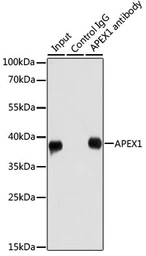
GTX35233 IP Image
IP analysis of HeLa cell lysate using GTX35233 APE1 antibody. Antibody amount : 1μg / 200μg lysate Dilution : 1:1000
|
|
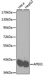
GTX35233 WB Image
WB analysis of various sample lysates using GTX35233 APE1 antibody. Loading : 25μg per lane
|
|
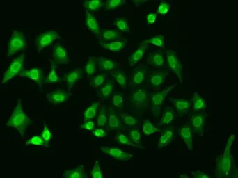
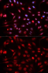
GTX35233 ICC/IF Image
ICC/IF analysis of HeLa cells using GTX35233 APE1 antibody. Blue : DAPI
|
|
GTX35233 WB Image
WB analysis of normal (control) and knockout (KO) 293T cell lysate using GTX35233 APE1 antibody. Dilution : 1:3000 Loading : 25μg per lane
|
|
GTX35233 ICC/IF Image
ICC/IF analysis of A549 cells using GTX35233 APE1 antibody.
|
REFERENCE
There are currently no references for APE1 antibody (GTX35233). Be the first to share your publications with this product.
REVIEW
There are currently no reviews for APE1 antibody (GTX35233). Be the first to share your experience with this product.