ARID2 antibody


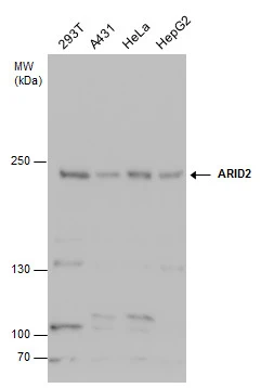
ARID2 antibody detects ARID2 protein by western blot analysis. Various whole cell extracts (30 μg) were separated by 5% SDS-PAGE, and the membrane was blotted with ARID2 antibody (GTX129443) diluted at 1:3000.
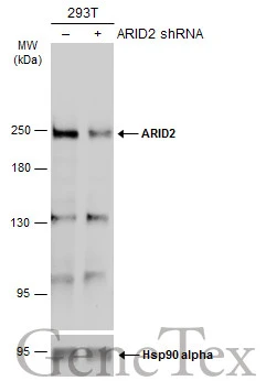
Non-transfected (–) and transfected (+) 293T whole cell extracts (50 μg) were separated by 5% SDS-PAGE, and the membrane was blotted with ARID2 antibody (GTX129443) diluted at 1:1000. The HRP-conjugated anti-rabbit IgG antibody (GTX213110-01) was used to detect the primary antibody, and the signal was developed with Trident ECL plus-Enhanced.
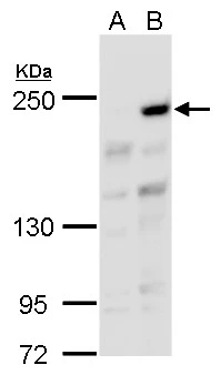
ARID2 antibody detects ARID2 protein by western blot analysis.
A. 30 μg 293T whole cell lysate/extract
B. 30 μg whole cell lysate/extract of human ARID2-transfected 293T cells
5 % SDS-PAGE
ARID2 antibody (GTX129443) dilution: 1:5000
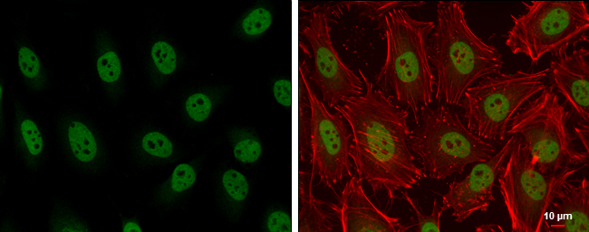
ARID2 antibody detects ARID2 protein at nucleus by immunofluorescent analysis.
Sample: HeLa cells were fixed in 4% paraformaldehyde at RT for 15 min.
Green: ARID2 protein stained by ARID2 antibody (GTX129443) diluted at 1:500.
Red: Phalloidin, a cytoskeleton marker, diluted at 1:200.
Scale bar = 10 μm.
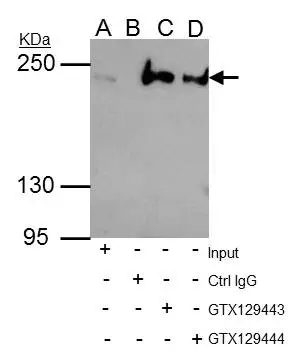
ARID2 antibody immunoprecipitates ARID2 protein in IP experiments.
IP samples: HeLa whole cell extract
A. 35 μg HeLa whole cell extract
B. Control with 4 μg of preimmune Rabbit IgG
C. Immunoprecipitation of ARID2 protein by 4 μg ARID2 antibody (GTX129443)
D. Immunoprecipitation of ARID2 protein by 4 μg ARID2 antibody (GTX129444)
5 % SDS-PAGE
The immunoprecipitated ARID2 protein was detected by ARID2 antibody (GTX129443) diluted at 1:3000.
[EasyBlot anti-rabbit IgG (GTX221666-01) was used as a secondary reagent]
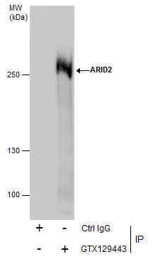
Immunoprecipitation of ARID2 protein from HepG2 whole cell extracts using 5 μg of ARID2 antibody (GTX129443).
Western blot analysis was performed using ARID2 antibody (GTX129443) diluted at 13000.
EasyBlot anti-Rabbit IgG (GTX221666-01) was used as a secondary reagent.
-
HostRabbit
-
ClonalityPolyclonal
-
IsotypeIgG
-
ApplicationsWB ICC/IF IP
-
ReactivityHuman