ARMET antibody


WB analysis of HEK293T cell or ARMET KO cell lysates using GTX31324 ARMET antibody.
Dilution : 1μg/mL
Loading : 10μg

IHC-P analysis of human brain tissue using GTX31324 ARMET antibody.
Working concentration : 2.5 μg/ml
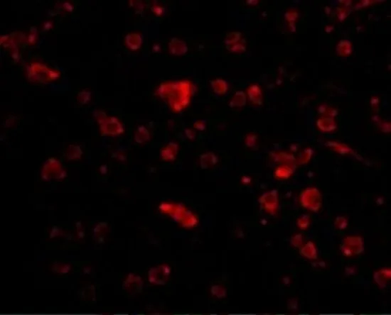
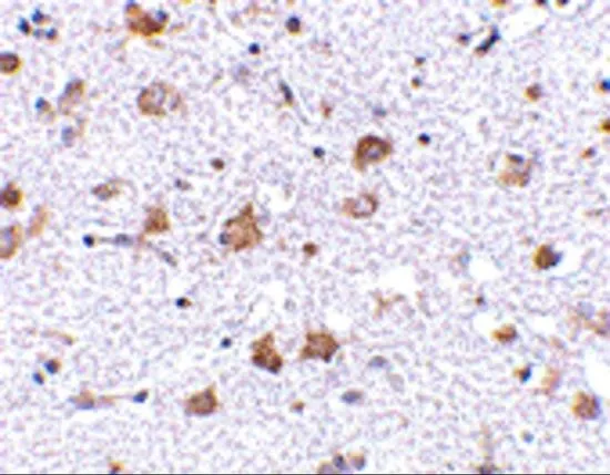
IHC-P analysis of human brain tissue using GTX31324 ARMET antibody.
Working concentration : 20 μg/ml
IHC-P analysis of human brain tissue using GTX31324 ARMET antibody.
Working concentration : 2.5 μg/ml
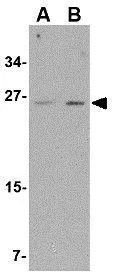
WB analysis of mouse brain tissue lysate using GTX31324 ARMET antibody.
Working concentration : (A) 1 and (B) 2 μg/ml
-
HostRabbit
-
ClonalityPolyclonal
-
IsotypeIgG
-
ApplicationsWB IHC-P ELISA
-
ReactivityHuman, Mouse, Rat