ARPC2 antibody
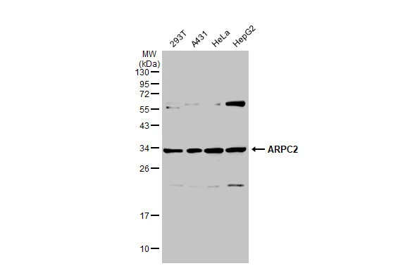
Various whole cell extracts (30 μg) were separated by 12% SDS-PAGE, and the membrane was blotted with ARPC2 antibody (GTX101984) diluted at 1:2000. The HRP-conjugated anti-rabbit IgG antibody (GTX213110-01) was used to detect the primary antibody.
ARPC2 antibody detects ARPC2 protein by western blot analysis.
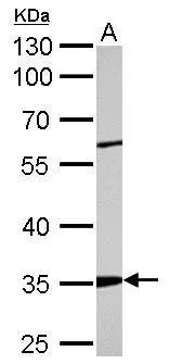
A. 50 μg rat kidney lysate/extract
10% SDS-PAGE
ARPC2 antibody (GTX101984) dilution: 1:5000
The HRP-conjugated anti-rabbit IgG antibody (GTX213110-01) was used to detect the primary antibody.
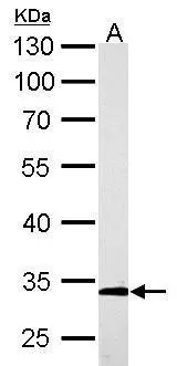
ARPC2 antibody detects ARPC2 protein by western blot analysis.
A. 50 μg mouse kidney lysate/extract
10% SDS-PAGE
ARPC2 antibody (GTX101984) dilution: 1:2000
The HRP-conjugated anti-rabbit IgG antibody (GTX213110-01) was used to detect the primary antibody.
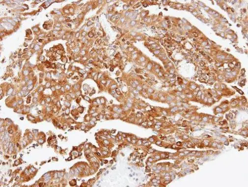
Immunohistochemical analysis of paraffin-embedded OVCA xenograft, using ARPC2(GTX101984) antibody at 1:100 dilution.
Antigen Retrieval: Trilogy™ (EDTA based, pH 8.0) buffer, 15min
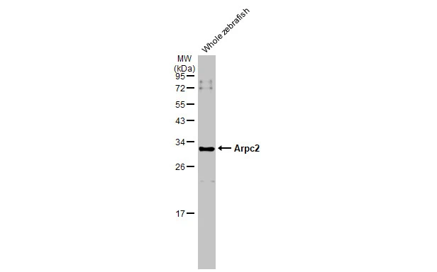
Whole cell extract (30 μg) was separated by 12% SDS-PAGE, and the membrane was blotted with ARPC2 antibody (GTX101984) diluted at 1:1000. The HRP-conjugated anti-rabbit IgG antibody (GTX213110-01) was used to detect the primary antibody.
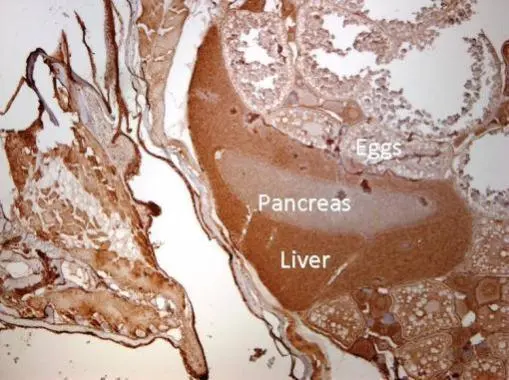
Immunohistochemical analysis of paraffin-embedded zebrafish tissue, using ARPC2 antibody (GTX101984) at 1:300 dilution.
-
HostRabbit
-
ClonalityPolyclonal
-
IsotypeIgG
-
ApplicationsWB IHC-P
-
ReactivityHuman, Mouse, Rat, Zebrafish