APPLICATION
Application Note
*Optimal dilutions/concentrations should be determined by the researcher.
Application |
Recommended Dilution |
1:500-1:3000 |
1:100-1:500 |
Not tested in other applications.
Calculated MW
Positive Control
293T , A431 , HeLa , HepG2
Predict Reactivity
Bovine, Pig, Rhesus Monkey(>80% identity)
PROPERTIES
Form
Liquid
Buffer
PBS, 20% Glycerol
Preservative
0.025% ProClin 300
Storage
Store as concentrated solution. Centrifuge briefly prior to opening vial. For short-term storage (1-2 weeks), store at 4ºC. For long-term storage, aliquot and store at -20ºC or below. Avoid multiple freeze-thaw cycles.
Concentration
1 mg/ml (Please refer to the vial label for the specific concentration.)
Antigen Species
Human
Immunogen
Carrier-protein conjugated synthetic peptide encompassing a sequence within the C-terminus region of human ATG4C. The exact sequence is proprietary.
Purification
Purified by antigen-affinity chromatography.
Conjugation
Unconjugated
RRID
AB_2886014
Note
For laboratory research use only. Not for any clinical, therapeutic, or diagnostic use in humans or animals. Not for animal or human consumption.
Purchasers shall not, and agree not to enable third parties to, analyze, copy, reverse engineer or otherwise attempt to determine the structure or sequence of the product.
TARGET
Synonyms
autophagy related 4C cysteine peptidase , APG4-C , APG4C , AUTL1 , AUTL3
Cellular Localization
Cytoplasm
Background
Autophagy is the process by which endogenous proteins and damaged organelles are destroyed intracellularly. Autophagy is postulated to be essential for cell homeostasis and cell remodeling during differentiation, metamorphosis, non-apoptotic cell death, and aging. Reduced levels of autophagy have been described in some malignant tumors, and a role for autophagy in controlling the unregulated cell growth linked to cancer has been proposed. This gene encodes a member of the autophagin protein family. The encoded protein is also designated as a member of the C-54 family of cysteine proteases. Alternate transcriptional splice variants, encoding the same protein, have been characterized. [provided by RefSeq]
Database
Research Area
DATA IMAGES
|
GTX129488 WB Image
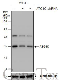
Non-transfected (–) and transfected (+) 293T whole cell extracts (30 μg) were separated by 10% SDS-PAGE, and the membrane was blotted with ATG4C antibody (GTX129488) diluted at 1:1000.
|
|
GTX129488 WB Image
ATG4C antibody detects ATG4C protein by western blot analysis. A. 30 μg 293T whole cell lysate/extract B. 30 μg A431 whole cell lysate/extract C. 30 μg HeLa whole cell lysate/extract D. 30 μg HepG2 whole cell lysate/extract 10 % SDS-PAGE ATG4C antibody (GTX129488) dilution: 1:1000
|
|
GTX129488 IP Image
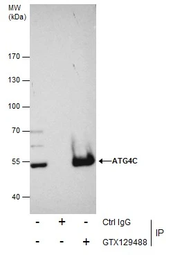
Immunoprecipitation of ATG4C protein from 293T whole cell extracts using 5 μg of ATG4C antibody (GTX129488). Western blot analysis was performed using ATG4C antibody (GTX129488). EasyBlot anti-Rabbit IgG (GTX221666-01) was used as a secondary reagent.
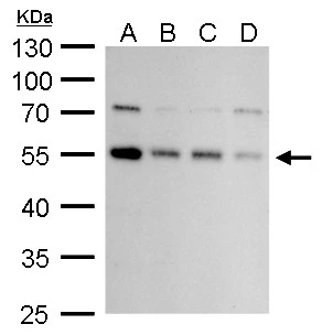
|
REFERENCE
There are currently no references for ATG4C antibody (GTX129488). Be the first to share your publications with this product.
REVIEW
There are currently no reviews for ATG4C antibody (GTX129488). Be the first to share your experience with this product.