ATP5B antibody, Internal
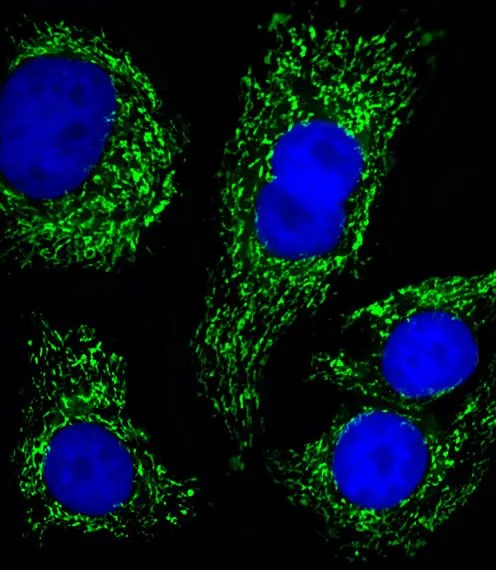
ICC/IF analysis of SK-Br-3 cells using GTX81939 ATP5B antibody, Internal.
Green : ATP5B
Blue : DAPI
Dilution : 1:25
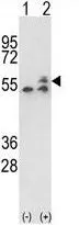
WB analysis of 293 cell lysate (2 ug/lane) either nontransfected (Lane 1) or transiently transfected with the ATP5B (Lane 2) using GTX81939 ATP5B antibody, Internal.
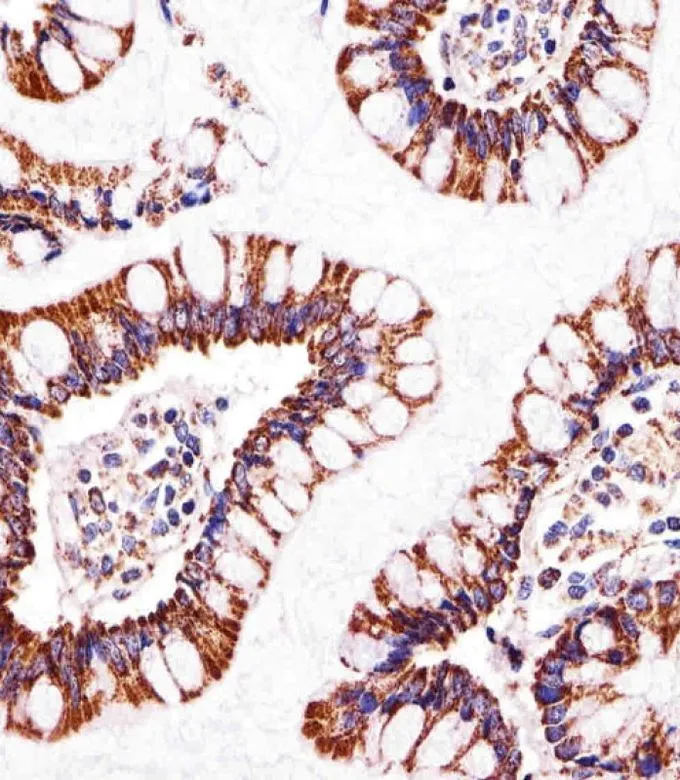
IHC-P analysis of human small intestine using GTX81939 ATP5B antibody, Internal.
Dilution : 1:25
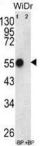
WB analysis of WiDr cell lysate using GTX81939 ATP5B antibody, Internal.
Lane 1 : pre-incubated without blocking peptide
Lane 2 : pre-incubate with blocking peptide
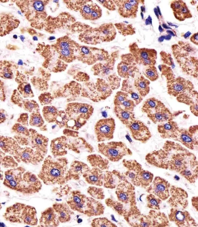
IHC-P analysis of human liver tissue using GTX81939 ATP5B antibody, Internal.
Dilution : 1:25
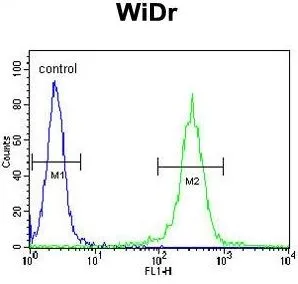
FACS analysis of WiDr cells using GTX81939 ATP5B antibody, Internal.
Green : primary antibody
Blue : negative control
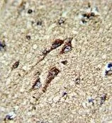
IHC-P analysis of human brain tissue using GTX81939 ATP5B antibody, Internal.
-
HostRabbit
-
ClonalityPolyclonal
-
IsotypeIgG
-
ApplicationsWB ICC/IF IHC-P FCM
-
ReactivityHuman