ATP6V1B2 antibody

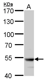
ATP6V1B2 antibody detects ATP6V1B2 protein by western blot analysis.
A. 50 μg rat brain lysate/extract
7.5 % SDS-PAGE
ATP6V1B2 antibody (GTX110783) dilution: 1:5000
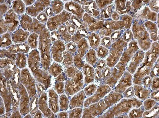
ATP6V1B2 antibody detects ATP6V1B2 protein at cytosol on mouse kidney by immunohistochemical analysis.
Sample: Paraffin-embedded mouse kidney.
ATP6V1B2 antibody (GTX110783) dilution: 1:500.
Antigen Retrieval: Trilogy™ (EDTA based, pH 8.0) buffer, 15min
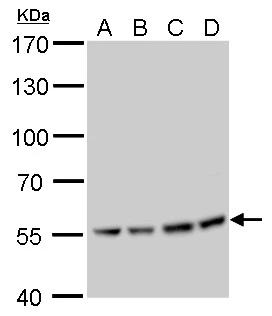
ATP6V1B2 antibody detects ATP6V1B2 protein by Western blot analysis.
A. 30 μg U87-MG whole cell lysate/extract
B. 30 μg SK-N-SH whole cell lysate/extract
C. 30 μg IMR32 whole cell lysate/extract
D. 30 μg SK-N-AS whole cell lysate/extract
7.5 % SDS-PAGE
ATP6V1B2 antibody (GTX110783) dilution: 1:1000
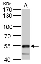
ATP6V1B2 antibody detects ATP6V1B2 protein by western blot analysis.
A. 50 μg mouse brain lysate/extract
7.5 % SDS-PAGE
ATP6V1B2 antibody (GTX110783) dilution: 1:5000
-
HostRabbit
-
ClonalityPolyclonal
-
IsotypeIgG
-
ApplicationsWB IHC-P
-
ReactivityHuman, Mouse, Rat