Bcl-XS antibody

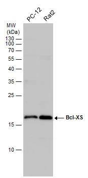
Various whole cell extracts (30 μg) were separated by 15% SDS-PAGE, and the membrane was blotted with Bcl-XS antibody (GTX124266) diluted at 1:1000.
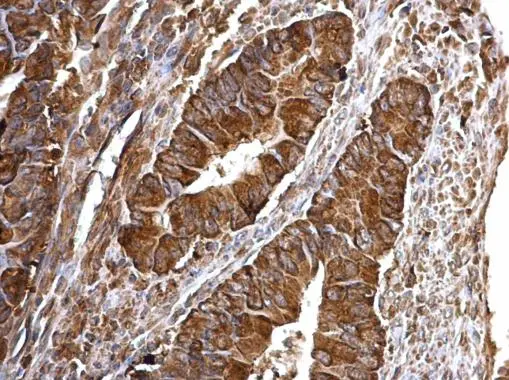
Bcl-XS antibody detects Bcl-XS protein at cytosol on human colon carcinoma by immunohistochemical analysis.
Sample: Paraffin-embedded human colon carcinoma.
Bcl-XS antibody (GTX124266) dilution: 1:500.
Antigen Retrieval: Trilogy™ (EDTA based, pH 8.0) buffer, 15min
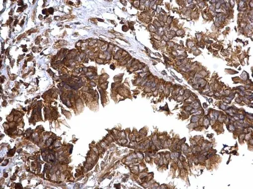
Bcl-XS antibody detects Bcl-XS protein at cytosol on human ovarian carcinoma by immunohistochemical analysis.
Sample: Paraffin-embedded human ovarian carcinoma.
Bcl-XS antibody (GTX124266) dilution: 1:500.
Antigen Retrieval: Trilogy™ (EDTA based, pH 8.0) buffer, 15min
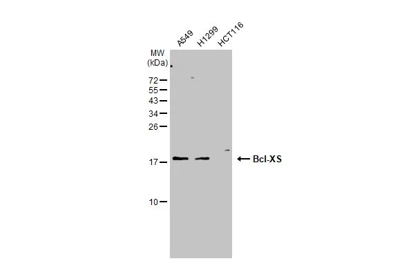
Various whole cell extracts (30 μg) were separated by 15% SDS-PAGE, and the membrane was blotted with Bcl-XS antibody (GTX124266) diluted at 1:1000. The HRP-conjugated anti-rabbit IgG antibody (GTX213110-01) was used to detect the primary antibody.
-
HostRabbit
-
ClonalityPolyclonal
-
IsotypeIgG
-
ApplicationsWB IHC-P IP
-
ReactivityHuman, Rat