APPLICATION
Application Note
*Optimal dilutions/concentrations should be determined by the researcher.
Application |
Recommended Dilution |
Assay dependent |
Assay dependent |
Assay dependent |
Assay dependent |
Assay dependent |
Assay dependent |
Not tested in other applications.
Calculated MW
Product Note
This antibody was raised against mouse BEST1 Extracellular domain.
PROPERTIES
Form
Liquid
Buffer
PBS, 1% BSA
Preservative
0.05% Sodium Azide
Storage
Store as concentrated solution. Centrifuge briefly prior to opening vial. For short-term storage (1-2 weeks), store at 4ºC. For long-term storage, aliquot and store at -20ºC or below. Avoid multiple freeze-thaw cycles.
Concentration
0.8 mg/ml (Please refer to the vial label for the specific concentration.)
Antigen Species
Mouse
Immunogen
Peptide (C)NPNKDYPGHEMD, corresponding to amino acid residues 259-270 of mouse Bestrophin-1 (Accession O88870) of 3rd extracellular loop
Purification
Purified by antigen-affinity chromatography
Conjugation
Unconjugated
Note
For laboratory research use only. Not for any clinical, therapeutic, or diagnostic use in humans or animals. Not for animal or human consumption.
Purchasers shall not, and agree not to enable third parties to, analyze, copy, reverse engineer or otherwise attempt to determine the structure or sequence of the product.
TARGET
Synonyms
ARB,BEST,BEST1,BMD,RP50,TU15B,VMD2,bestrophin 1,Bestrophin 1
Cellular Localization
Cell membrane,Basolateral cell membrane
Background
This gene encodes a member of the bestrophin gene family. This small gene family is characterized by proteins with a highly conserved N-terminus with four to six transmembrane domains. Bestrophins may form chloride ion channels or may regulate voltage-gated L-type calcium-ion channels. Bestrophins are generally believed to form calcium-activated chloride-ion channels in epithelial cells but they have also been shown to be highly permeable to bicarbonate ion transport in retinal tissue. Mutations in this gene are responsible for juvenile-onset vitelliform macular dystrophy (VMD2), also known as Best macular dystrophy, in addition to adult-onset vitelliform macular dystrophy (AVMD) and other retinopathies. Alternative splicing results in multiple variants encoding distinct isoforms.[provided by RefSeq, Nov 2008]
Database
Research Area
DATA IMAGES
|
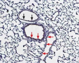
GTX04928 IHC-P Image
IHC-P analysis of rat lung tissue using GTX04928 Bestrophin 1 antibody. BEST1 is expressed both in the respiratory epithelium (red arrows) and in vascular smooth muscle (black arrows). Dilution : 1:100
|
|
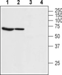
GTX04928 WB Image
WB analysis of various samples using GTX04928 Bestrophin 1 antibody prior incubated with (Lane 3-4) or without (Lane 1-2) blocking peptide. Lane 1 and Lane 3: rat lung tissue lysate Lane 2 and lane 4 : rat eye tissue lysate Dilution : 1:200
|
|
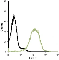
GTX04928 FACS Image
FACS analysis of live intact Jurkat cells using GTX04928 Bestrophin 1 antibody. Green line : Primary antibody Black line : Control Dilution : 1:20
|
REFERENCE
There are currently no references for Bestrophin 1 antibody (GTX04928). Be the first to share your publications with this product.
REVIEW
There are currently no reviews for Bestrophin 1 antibody (GTX04928). Be the first to share your experience with this product.