Bestrophin 1 antibody
Cat. No. GTX108854
Cat. No. GTX108854
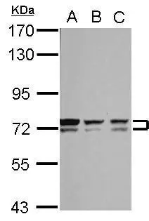
GTX108854 WB Image
Sample (30 ug of whole cell lysate)
A: NT2D1
B: PC-3
C: U87-MG
7.5% SDS PAGE
GTX108854 diluted at 1:1000
1 / 2
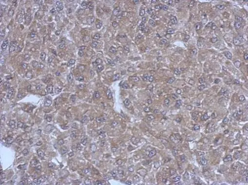
GTX108854 IHC-P Image
Immunohistochemical analysis of paraffin-embedded U87 xenograft, using Bestrophin 1(GTX108854) antibody at 1:500 dilution.
Antigen Retrieval: Trilogy™ (EDTA based, pH 8.0) buffer, 15min
2 / 2
-
HostRabbit
-
ClonalityPolyclonal
-
IsotypeIgG
-
ApplicationsWB IHC-P
-
ReactivityHuman