Bmi1 antibody


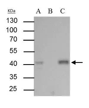
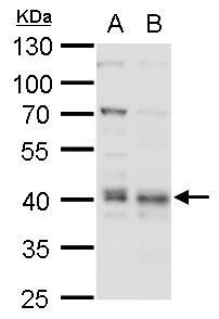
Bmi1 antibody immunoprecipitates Bmi1 protein in IP experiments. IP Sample: 293T whole cell lysate/extract A : 30 μg whole cell lysate/extract of Bmi1 protein expressing 293T cells B : Control with 2.5 μg of pre-immune rabbit IgG C : Immunoprecipitation of Bmi1 protein by 2.5 μg of Bmi1 antibody (GTX114008) 10% SDS-PAGE The immunoprecipitated Bmi1 protein was detected by Bmi1 antibody (GTX114008) diluted at 1 : 1000. EasyBlot anti-rabbit IgG (HRP) (GTX221666-01) was used as a secondary reagent.
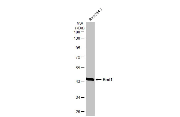
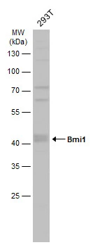
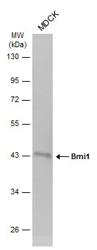
Whole cell extract (30 μg) was separated by 10% SDS-PAGE, and the membrane was blotted with Bmi1 antibody (GTX114008) diluted at 1:1000. The HRP-conjugated anti-rabbit IgG antibody (GTX213110-01) was used to detect the primary antibody.
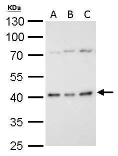
Bmi1 antibody detects BMI1 protein by western blot analysis.
A. 30 μg C8D30 whole cell lysate/extract
B. 30 μg BCL-1 whole cell lysate/extract
C. 30 μg Raw264.7 whole cell lysate/extract
10% SDS-PAGE
Bmi1 antibody (GTX114008) dilution: 1:1000
The HRP-conjugated anti-rabbit IgG antibody (GTX213110-01) was used to detect the primary antibody.
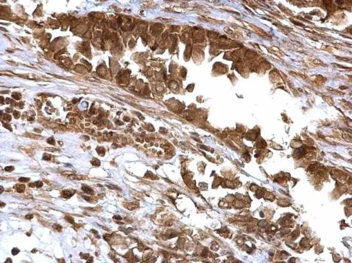
Bmi1 antibody detects Bmi1 protein at cytosol and nucleus on human ovarian carcinoma by immunohistochemical analysis.
Sample: Paraffin-embedded human ovarian carcinoma.
Bmi1 antibody (GTX114008) dilution: 1:500.
Antigen Retrieval: Trilogy™ (EDTA based, pH 8.0) buffer, 15min
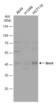
Various whole cell extracts (30 μg) were separated by 10% SDS-PAGE, and the membrane was blotted with Bmi1 antibody (GTX114008) diluted at 1:1000. The HRP-conjugated anti-rabbit IgG antibody (GTX213110-01) was used to detect the primary antibody.
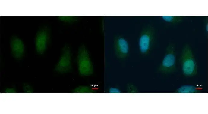
BMI1 antibody [N1C3-2] detects BMI1 protein by immunofluorescent analysis.
Sample: HeLa cells were fixed in 4% paraformaldehyde for 15 min.
Green: BMI1 protein stained by BMI1 antibody (GTX114008) diluted at 1:1000.
Blue: Hoechst 33342 staining.
Scale bar = 10 μm.
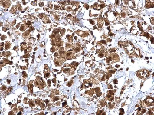
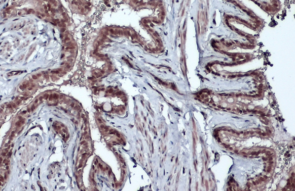
Bmi1 antibody detects Bmi1 protein at cytosol and nucleus on human breast carcinoma by immunohistochemical analysis.
Sample: Paraffin-embedded human breast carcinoma.
Bmi1 antibody (GTX114008) dilution: 1:500.
Antigen Retrieval: Trilogy™ (EDTA based, pH 8.0) buffer, 15min
Bmi1 antibody detects BMI1 protein by western blot analysis.
A. 30 μg THP-1 whole cell lysate/extract
B. 30 μg HL-60 whole cell lysate/extract
10% SDS-PAGE
Bmi1 antibody (GTX114008) dilution: 1:1000
The HRP-conjugated anti-rabbit IgG antibody (GTX213110-01) was used to detect the primary antibody.
Bmi1 antibody detects Bmi1 protein by western blot analysis.
A. 30 μg K562 whole cell lysate/extract
B. 30 μg THP-1 whole cell lysate/extract
10% SDS-PAGE
Bmi1 antibody (GTX114008) dilution: 1:1000
The HRP-conjugated anti-rabbit IgG antibody (GTX213110-01) was used to detect the primary antibody.
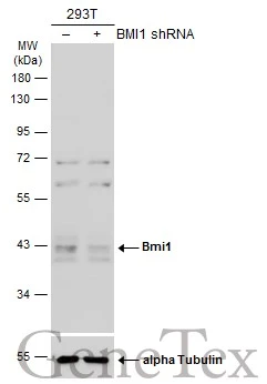
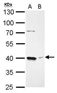
Non-transfected (–) and transfected (+) HeLa whole cell extracts (30 μg) were separated by 10% SDS-PAGE, and the membrane was blotted with Bmi1 antibody (GTX114008) diluted at 1:500. The HRP-conjugated anti-rabbit IgG antibody (GTX213110-01) was used to detect the primary antibody.
Whole cell extract (30 μg) was separated by 10% SDS-PAGE, and the membrane was blotted with Bmi1 antibody (GTX114008) diluted at 1:1000. The HRP-conjugated anti-rabbit IgG antibody (GTX213110-01) was used to detect the primary antibody.
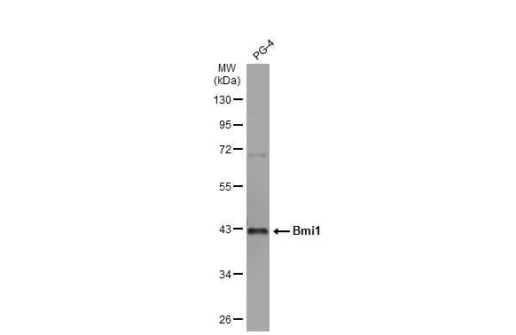
Whole cell extract (30 μg) was separated by 10% SDS-PAGE, and the membrane was blotted with Bmi1 antibody (GTX114008) diluted at 1:1000. The HRP-conjugated anti-rabbit IgG antibody (GTX213110-01) was used to detect the primary antibody.
Whole cell extract (30 μg) was separated by 10% SDS-PAGE, and the membrane was blotted with Bmi1 antibody (GTX114008) diluted at 1:1000. The HRP-conjugated anti-rabbit IgG antibody (GTX213110-01) was used to detect the primary antibody, and the signal was developed with Trident ECL plus-Enhanced.
Bmi1 antibody detects Bmi1 protein at cytoplasm and nucleus by immunohistochemical analysis.Sample: Paraffin-embedded dog lung.Bmi1 stained by Bmi1 antibody (GTX114008) diluted at 1:500.Antigen Retrieval: Citrate buffer, pH 6.0, 15 min
Bmi1 antibody detects Bmi1 protein at nucleus by immunofluorescent analysis.Sample: PG-4 cells were fixed in 4% paraformaldehyde at RT for 15 min.Green: Bmi1 stained by Bmi1 antibody (GTX114008) diluted at 1:1000.Blue: Hoechst 33342 staining.Scale bar= 10 μm.

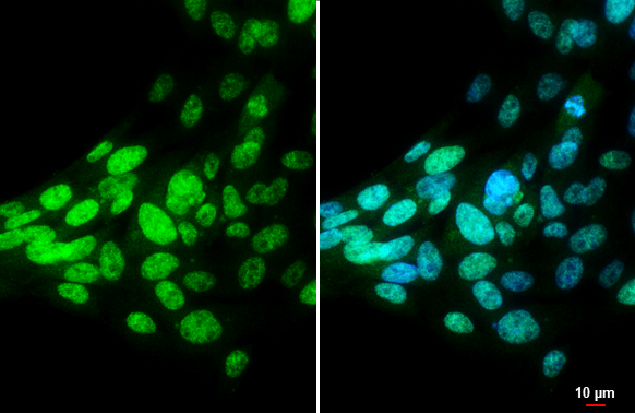
Bmi1 antibody detects Bmi1 protein at nucleus by immunofluorescent analysis.Sample: MDCK cells were fixed in 4% paraformaldehyde at RT for 15 min.Green: Bmi1 stained by Bmi1 antibody (GTX114008) diluted at 1:1000.Red: phalloidin, a cytoskeleton marker, diluted at 1:100.Scale bar= 10 μm.
-
HostRabbit
-
ClonalityPolyclonal
-
IsotypeIgG
-
ApplicationsWB ICC/IF IHC-P IP
-
ReactivityHuman, Mouse, Cat, Dog