Bmi1 antibody
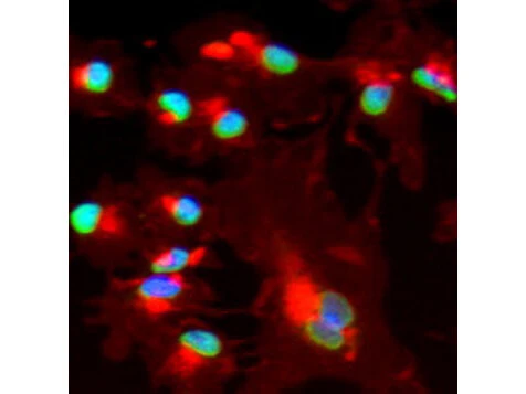
ICC/IF analysis of MeOH-fixed HepG2 cells using GTX45791 Bmi1 antibody.
Green : Primary antibody
Red : Wheat germ agglutinin
Blue : DAPI
Dilution : 1:200
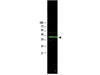
WB analysis of U2OS whole cell lysate using GTX45791 Bmi1 antibody.
Loading : 20 μg
Dilution : 1:1000
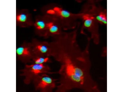
Immunofluorescence using affinity purified goat anti Bmi1(GTX45791) shows nuclear staining (green) of methanol fixed (100%, 5 min) HepG2 cells. The cells were blocked and permeabilized in 1%BSA / 10% normal donkey serum / 0.3M glycine in 0.1% PBS-Tween for 1h prior to incubation with the primary antibody (1:200 dilution) overnight at +4ºC and detected with a 488nm fluorescent dye conjugated secondary Ab. Cell nuclei are stained with DAPI (blue) and plasma membranes are stained with WGA (red).
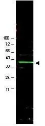
Western blot using GeneTex's Affinity Purified anti-Bmi1 antibody shows detection of a band ~37 kDa corresponding to human Bmi1 (arrowhead). Approximately 20 μg of a U2OS whole cell lysate was separated by 4-20% SDS-PAGE and transferred onto nitrocellulose. After blocking in PBS containing 5% nonfat dry milk, the membrane was probed overnight at 4º C with the primary antibody diluted to 1:1,000 in PBS containing 1% nonfat dry milk. The membrane was washed and reacted with a 1:20,000 dilution of IRDye800 conjugated rabbit anti-Goat IgG [H&L] for 45 min at room temperature. IRDye800 fluorescence image was captured using the OdysseyR Infrared Imaging System developed by LI-COR. IRDye is a trademark of LI-COR, Inc. Other detection systems will yield similar results.
-
HostGoat
-
ClonalityPolyclonal
-
IsotypeIgG
-
ApplicationsWB ICC/IF ELISA Multiplexing
-
ReactivityHuman