CASK antibody
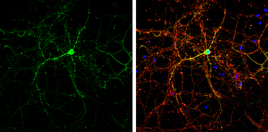
CASK antibody detects CASK protein by immunofluorescent analysis.
Sample: DIV14 rat E18 primary cortical neurons were fixed in 4% paraformaldehyde at RT for 15 min.
Green: CASK protein stained by CASK antibody (GTX133275) diluted at 1:500.
Red: beta Tubulin 3/ Tuj1, stained by beta Tubulin 3/ Tuj1 antibody [GT1338] (GTX631831) diluted at 1:500.
Blue: Fluoroshield with DAPI (GTX30920).
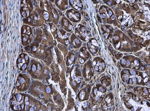
CASK antibody detects CASK protein at cytoplasm in mouse colon by immunohistochemical analysis.
Sample: Paraffin-embedded mouse colon.
CASK antibody (GTX133275) diluted at 1:500.
Antigen Retrieval: Citrate buffer, pH 6.0, 15 min
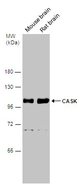
Various tissue extracts (50 μg) were separated by 7.5% SDS-PAGE, and the membrane was blotted with CASK antibody (GTX133275) diluted at 1:5000.
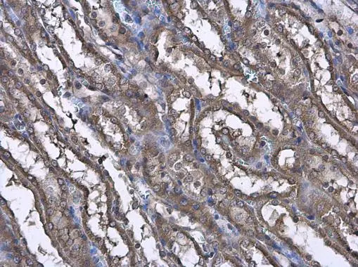
CASK antibody detects CASK protein at cytoplasm in rat kidney by immunohistochemical analysis.
Sample: Paraffin-embedded rat kidney.
CASK antibody (GTX133275) diluted at 1:500.
Antigen Retrieval: Citrate buffer, pH 6.0, 15 min
-
HostRabbit
-
ClonalityPolyclonal
-
IsotypeIgG
-
ApplicationsWB ICC/IF IHC-P
-
ReactivityMouse, Rat