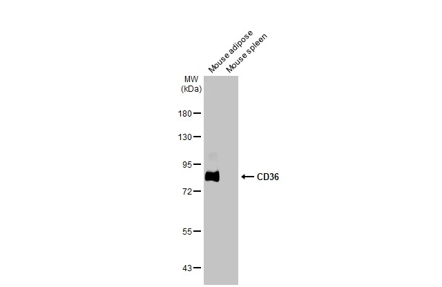
APPLICATION
Application Note
*Optimal dilutions/concentrations should be determined by the researcher.
Application |
Dilution |
1:500-1:3000 |
1:100-1:1000 |
Not tested in other applications.
Calculated MW
Positive Control
HeLa , HepG2 , Mouse brown adipose
Predict Reactivity
Rat, Bovine, Dog, Pig, Rhesus Monkey(>80% identity)
PROPERTIES
Form
Liquid
Buffer
0.1M Tris-Glycine (pH7), 20% Glycerol
Preservative
0.01% Thimerosal
Storage
Store as concentrated solution. Centrifuge briefly prior to opening vial. For short-term storage (1-2 weeks), store at 4ºC. For long-term storage, aliquot and store at -20ºC or below. Avoid multiple freeze-thaw cycles.
Concentration
0.71 mg/ml (Please refer to the vial label for the specific concentration.)
Antigen Species
Human
Immunogen
Recombinant protein encompassing a sequence within the center region of human CD36. The exact sequence is proprietary.
Purification
Purified by antigen-affinity chromatography.
Conjugation
Unconjugated
RRID
AB_10731492
Note
For laboratory use only. Not for any clinical, therapeutic, or diagnostic use in humans or animals. Not for animal or human consumption.
TARGET
Synonyms
CD36 molecule , BDPLT10 , CHDS7 , FAT , GP3B , GP4 , GPIV , PASIV , SCARB3
Cellular Localization
Membrane
Background
The protein encoded by this gene is the fourth major glycoprotein of the platelet surface and serves as a receptor for thrombospondin in platelets and various cell lines. Since thrombospondins are widely distributed proteins involved in a variety of adhesive processes, this protein may have important functions as a cell adhesion molecule. It binds to collagen, thrombospondin, anionic phospholipids and oxidized LDL. It directly mediates cytoadherence of Plasmodium falciparum parasitized erythrocytes and it binds long chain fatty acids and may function in the transport and/or as a regulator of fatty acid transport. Mutations in this gene cause platelet glycoprotein deficiency. Multiple alternatively spliced transcript variants encoding the same protein have been found for this gene. [provided by RefSeq]
Database
Research Area
DATA IMAGES
|
GTX112891 WB Image
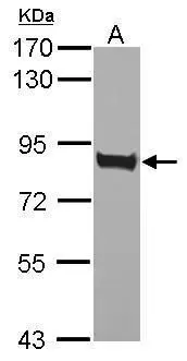
Sample (50 ug of whole cell lysate) A: Mouse brown adipose 7.5% SDS PAGE GTX112891 diluted at 1:1000
|
|
GTX112891 IHC-P Image
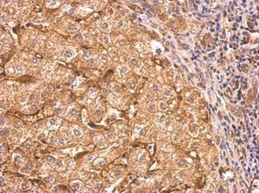
CD36 antibody detects CD36 protein at cytosol and weak membrane on human hepatoma by immunohistochemical analysis. Sample: Paraffin-embedded hepatoma. CD36 antibody (GTX112891) dilution: 1:500.
Antigen Retrieval: Trilogy™ (EDTA based, pH 8.0) buffer, 15min
|
|
GTX112891 WB Image
Sample (30 ug of whole cell lysate) A: HeLa B: HepG2 7.5% SDS PAGE GTX112891 diluted at 1:2000
|
|
GTX112891 IHC-P Image
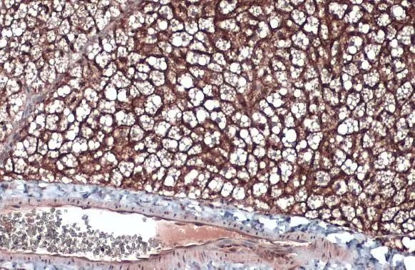
CD36 antibody detects CD36 protein by immunohistochemical analysis.Sample: Paraffin-embedded mouse brown adipocyte.CD36 stained by CD36 antibody (GTX112891) diluted at 1:500.Antigen Retrieval: Citrate buffer, pH 6.0, 15 min
|
|
GTX112891 WB Image
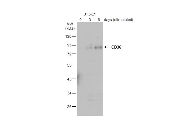
Unstimulatd and stimulatd 3T3-L1 whole cell extracts (20 μg) were separated by 10% SDS-PAGE, and the membrane was blotted with CD36 antibody (GTX112891) diluted at 1:2000. The HRP-conjugated anti-rabbit IgG antibody (GTX213110-01) was used to detect the primary antibody, and the signal was developed with Trident ECL plus-Enhanced. (The differentiation stimulated medium is composed by basal medium, 10% FBS, 50 ug/ml gentamicin, 1 nM L-glutamin, 500 uM IBMX, 1 uM dexamethasone, 2 uM rosiglitazone and 1 ug/ml insulin.)
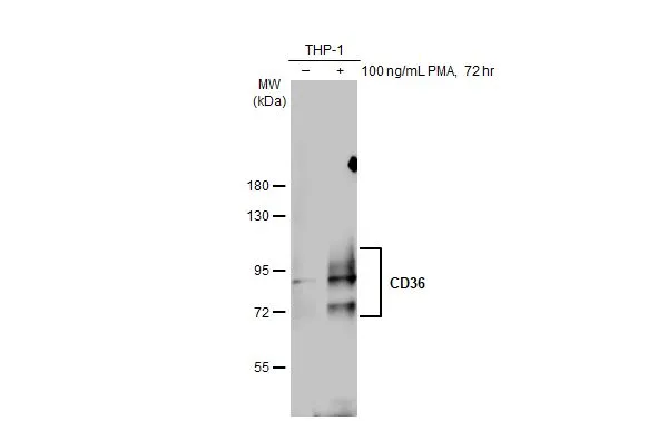
|
REFERENCE
There are currently no references for CD36 antibody (GTX112891). Be the first to share your publications with this product.
REVIEW
There are currently no reviews for CD36 antibody (GTX112891). Be the first to share your experience with this product.