CD44 antibody


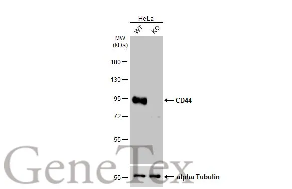
Non-transfected (–) and transfected (+) Wild-type (WT) and G CD44 knockout (KO) HeLa cell extracts (30 μg) were separated by 7.5% SDS-PAGE, and the membrane was blotted with CD44 antibody (GTX102111) diluted at 1:7000. The HRP-conjugated anti-rabbit IgG antibody (GTX213110-01) was used to detect the primary antibody.
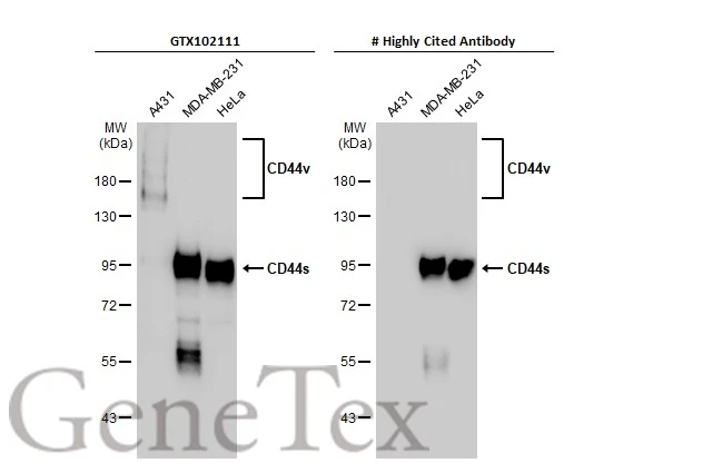
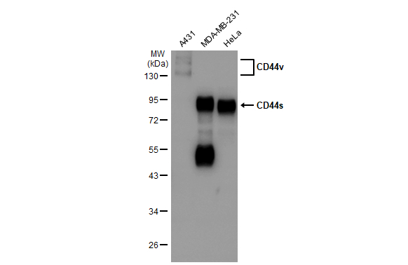
Various whole cell extracts (30 μg) were separated by 7.5% SDS-PAGE, and the membranes were blotted with CD44 antibody (GTX102111) diluted at 1:5000 and competitor's antibody (# Highly Cited Antibody) diluted at 1:5000. The HRP-conjugated anti-rabbit IgG antibody (GTX213110-01) was used to detect the primary antibody.
*The competitor is not affiliated with GeneTex and does not endorse this product.
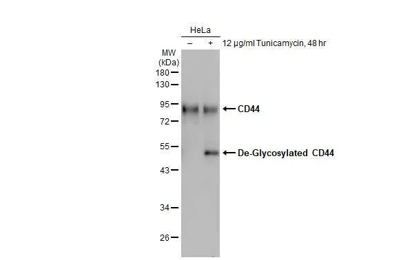
The observed M.W. is different from the predicted size. It is possibly due to post-translational modifications.
Reference: PMID: 10050880
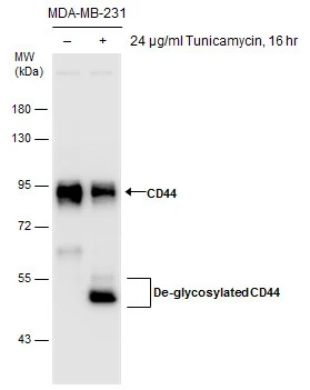
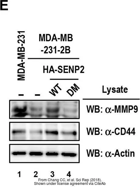
Untreated (–) and treated (+) MDA-MB-231 whole cell extracts (30 μg) were separated by 7.5% SDS-PAGE, and the membrane was blotted with CD44 antibody (GTX102111) diluted at 1:7000. The HRP-conjugated anti-rabbit IgG antibody (GTX213110-01) was used to detect the primary antibody.
The observed M.W. is different from the predicted size. It is possibly due to post-translational modifications.
Reference: PMID: 10050880
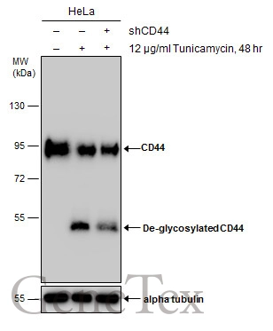
Untreated (–) and treated (+) HeLa whole cell extracts (30 μg) were separated by 7.5% SDS-PAGE, and the membrane was blotted with CD44 antibody (GTX102111) diluted at 1:10000. The HRP-conjugated anti-rabbit IgG antibody (GTX213110-01) was used to detect the primary antibody.
Untreated (–) and treated (+) HeLa whole cell extracts (30 μg) were separated by 10% SDS-PAGE, and the membrane was blotted with CD44 antibody (GTX102111) diluted at 1:45000. The HRP-conjugated anti-rabbit IgG antibody (GTX213110-01) was used to detect the primary antibody, and the signal was developed with Trident ECL plus-Enhanced.
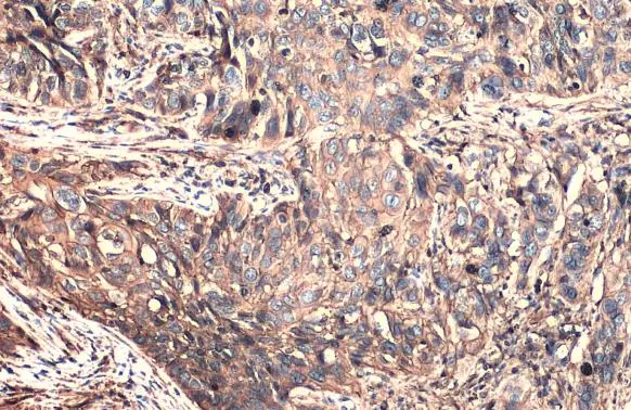
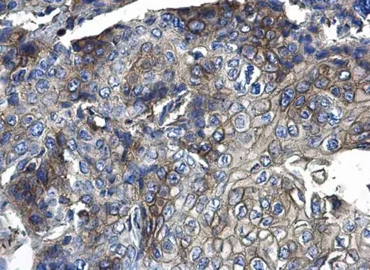
CD44 antibody detects CD44 protein at cell membrane and cytoplasm by immunohistochemical analysis.Sample: Paraffin-embedded human cervical carcinoma.CD44 stained by CD44 antibody (GTX102111) diluted at 1:500.Antigen Retrieval: Citrate buffer, pH 6.0, 15 min
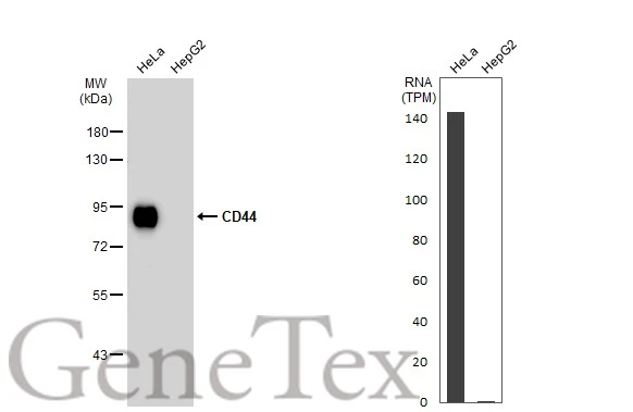
Various whole cell extracts (30 μg) were separated by 7.5% SDS-PAGE, and the membrane was blotted with CD44 antibody (GTX102111) diluted at 1:5000. The HRP-conjugated anti-rabbit IgG antibody (GTX213110-01) was used to detect the primary antibody. Corresponding RNA expression data for the same cell lines are based on Human Protein Atlas program.
Immunohistochemical analysis of paraffin-embedded Human pancreatic tumor, using CD44(GTX102111) antibody at 1:100 dilution.
Antigen Retrieval: Trilogy™ (EDTA based, pH 8.0) buffer, 15min
Various whole cell extracts (30 μg) were separated by 10% SDS-PAGE, and the membrane was blotted with CD44 antibody (GTX102111) diluted at 1:5000. The HRP-conjugated anti-rabbit IgG antibody (GTX213110-01) was used to detect the primary antibody.
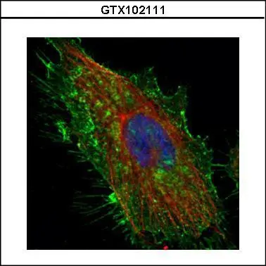
Confocal immunofluorescence analysis (Olympus FV10i) of methanol-fixed HeLa, using CD44(GTX102111) antibody (Green) at 1:500 dilution. Alpha-tubulin filaments were labeled with GTX11304 (Red) at 1:2000.
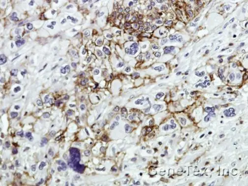
CD44 antibody detects CD44 protein at membrane and cytoplasm on human endometrial carcinoma by immunohistochemical analysis.
Sample: Paraffin-embedded human endometrial carcinoma .
CD44 antibody (GTX102111) diluted at 1:500.
Antigen Retrieval: Trilogy™ (EDTA based, pH 8.0) buffer, 15min
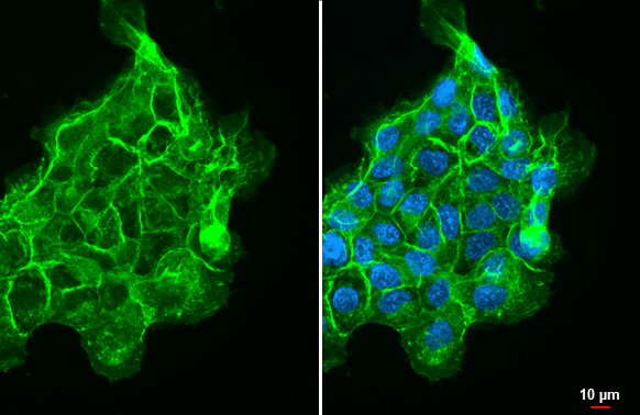
CD44 antibody detects CD44 protein at cell membrane by immunofluorescent analysis.Sample: A431 cells were fixed in ice-cold MeOH for 5 min.Green: CD44 stained by CD44 antibody (GTX102111) diluted at 1:500.Blue: Fluoroshield with DAPI (GTX30920).

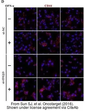
The data was published in the journal Oncotarget in 2016. PMID: 27487131
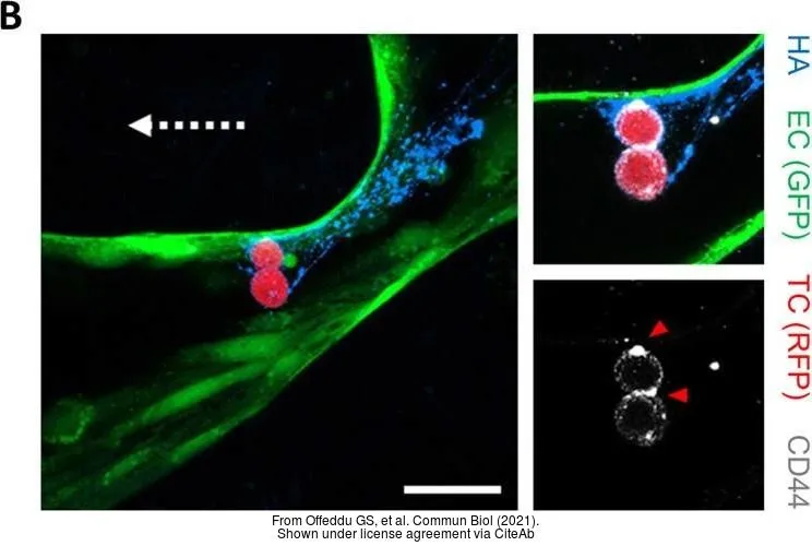
The data was published in the 2021 in Commun Biol. PMID: 33637851
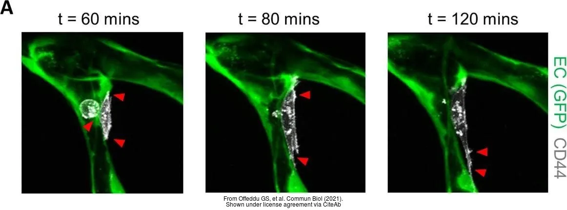
The data was published in the 2021 in Commun Biol. PMID: 33637851
-
HostRabbit
-
ClonalityPolyclonal
-
IsotypeIgG
-
ApplicationsWB ICC/IF IHC-P IHC-Fr IP LCI
-
ReactivityHuman, Rat, Rabbit