APPLICATION
Application Note
*Optimal dilutions/concentrations should be determined by the researcher.
Application |
Recommended Dilution |
1:500-1:3000 |
Assay dependent |
Not tested in other applications.
Calculated MW
Positive Control
H1299 , HeLa
Product Note
IP/MS validation was supported by references (PMID:30377419)
Predict Reactivity
Bovine, Dog(>80% identity)
PROPERTIES
Form
Liquid
Buffer
PBS, 20% Glycerol
Preservative
0.025% ProClin 300
Storage
Store as concentrated solution. Centrifuge briefly prior to opening vial. For short-term storage (1-2 weeks), store at 4ºC. For long-term storage, aliquot and store at -20ºC or below. Avoid multiple freeze-thaw cycles.
Concentration
0.1 mg/ml (Please refer to the vial label for the specific concentration.)
Antigen Species
Human
Immunogen
Recombinant protein encompassing a sequence within the Intracellular domain of human CD44. The exact sequence is proprietary.
Purification
Purified by antigen-affinity chromatography.
Conjugation
Unconjugated
RRID
AB_2886513
Note
For laboratory research use only. Not for any clinical, therapeutic, or diagnostic use in humans or animals. Not for animal or human consumption.
Purchasers shall not, and agree not to enable third parties to, analyze, copy, reverse engineer or otherwise attempt to determine the structure or sequence of the product.
TARGET
Synonyms
CD44 molecule (Indian blood group) , CDW44 , CSPG8 , ECMR-III , HCELL , HUTCH-I , IN , LHR , MC56 , MDU2 , MDU3 , MIC4 , Pgp1
Cellular Localization
Membrane
Background
The protein encoded by this gene is a cell-surface glycoprotein involved in cell-cell interactions, cell adhesion and migration. It is a receptor for hyaluronic acid (HA) and can also interact with other ligands, such as osteopontin, collagens, and matrix metalloproteinases (MMPs). This protein participates in a wide variety of cellular functions including lymphocyte activation, recirculation and homing, hematopoiesis, and tumor metastasis. Transcripts for this gene undergo complex alternative splicing that results in many functionally distinct isoforms, however, the full length nature of some of these variants has not been determined. Alternative splicing is the basis for the structural and functional diversity of this protein, and may be related to tumor metastasis. [provided by RefSeq]
Database
Research Area
DATA IMAGES
|
GTX131669 WB Image
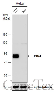
Wild-type (WT) and CD44 knockout (KO) HeLa cell extracts (30 μg) were separated by 7.5% SDS-PAGE, and the membrane was blotted with CD44 antibody (GTX131669) diluted at 1:1000.
|
|
GTX131669 WB Image
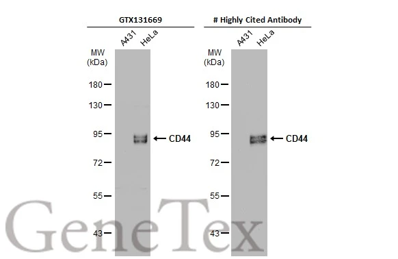
Various whole cell extracts (30 μg) were separated by 7.5% SDS-PAGE, and the membranes were blotted with CD44 antibody (GTX131669) diluted at 1:500 and competitor's antibody diluted at 1:500. The HRP-conjugated anti-rabbit IgG antibody (GTX213110-01) was used to detect the primary antibody.
*The competitor is not affiliated with GeneTex and does not endorse this product.
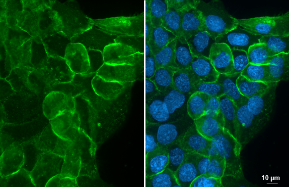
|
|
GTX131669 WB Image
CD44 antibody detects CD44 protein by western blot analysis. Whole cell extracts (30 μg) was separated by 7.5% SDS-PAGE, and the membrane was blotted with CD44 antibody (GTX131669) at a dilution of 1:1000.
|
|
GTX131669 WB Image
Whole cell extract (30 μg) was separated by 7.5% SDS-PAGE, and the membrane was blotted with CD44 antibody (GTX131669) diluted at 1:1000. The HRP-conjugated anti-rabbit IgG antibody (GTX213110-01) was used to detect the primary antibody.
|
|
GTX131669 WB Image
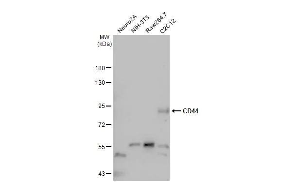
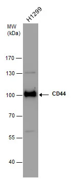
Various whole cell extracts (30 μg) were separated by 7.5% SDS-PAGE, and the membrane was blotted with CD44 antibody (GTX131669) diluted at 1:1000. The HRP-conjugated anti-rabbit IgG antibody (GTX213110-01) was used to detect the primary antibody, and the signal was developed with Trident ECL plus-Enhanced.
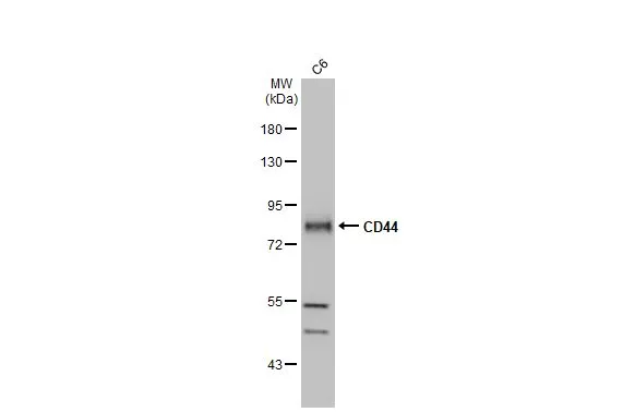
|
REFERENCE
REVIEW
There are currently no reviews for CD44 antibody (GTX131669). Be the first to share your experience with this product.