CD63 antibody

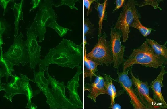
CD63 antibody detects CD63 protein at cell membrane by immunofluorescent analysis.Sample: HeLa cells were fixed in ice-cold MeOH for 5 min.Green: CD63 stained by CD63 antibody (GTX135220) diluted at 1:1000.Red: alpha Tubulin, a cytoskeleton marker, stained by alpha Tubulin antibody [GT114] (GTX628802) diluted at 1:1000.Blue: Fluoroshield with DAPI (GTX30920).
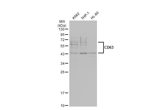
Various whole cell extracts (30 μg) were separated by 12% SDS-PAGE, and the membrane was blotted with CD63 antibody (GTX135220) diluted at 1:1000. The HRP-conjugated anti-rabbit IgG antibody (GTX213110-01) was used to detect the primary antibody.
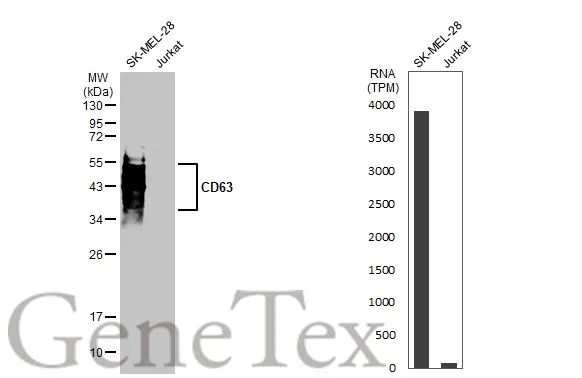
Various whole cell extracts (30 μg) were separated by 12% SDS-PAGE, and the membrane was blotted with CD63 antibody (GTX135220) diluted at 1:1000. The HRP-conjated anti-rabbit IgG antibody (GTX213110-01) was used to detect the primary antibody. Corresponding RNA expression data for the same cell lines are based on Human Protein Atlas program.
-
HostRabbit
-
ClonalityPolyclonal
-
IsotypeIgG
-
ApplicationsWB ICC/IF
-
ReactivityHuman