CD81 antibody

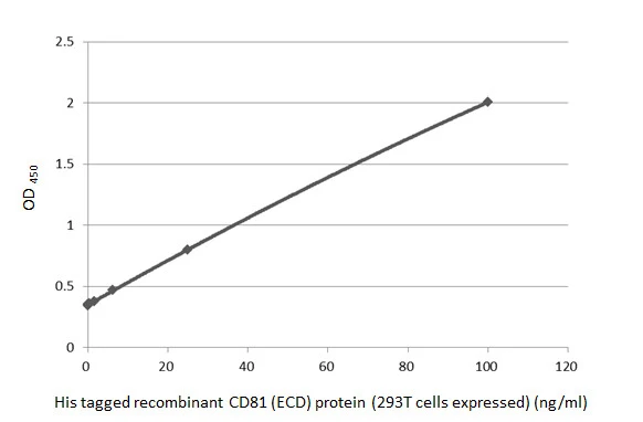
Sandwich ELISA detection of His tagged recombinant CD81 (ECD) protein (293T cells expressed) using antibodies as below. Capture: CD81 antibody (GTX135297) (5 μg/mL) Detection: HRP-conjugated CD81 antibody [HL4048] (GTX642463) (1 μg/mL)Please notice that GTX642463 needs to be conjugated to HRP to function as the detection antibody when paired with GTX135297. Please contact us for custom HRP-conjugated antibody.
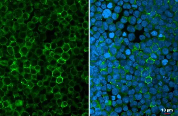
CD81 antibody detects CD81 protein at cell membrane by immunofluorescent analysis.
Sample: Jurkat cells were fixed in 4% paraformaldehyde at RT for 15 min.
Green: CD81 stained by CD81 antibody (GTX135297) diluted at 1:500.
Blue: Fluoroshield with DAPI (GTX30920).
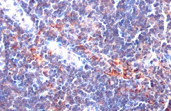
CD81 antibody detects CD81 protein at cell membrane by immunohistochemical analysis.Sample: Paraffin-embedded mouse lymph node.CD81 stained by CD81 antibody (GTX135297) diluted at 1:500.Antigen Retrieval: Citrate buffer, pH 6.0, 15 min
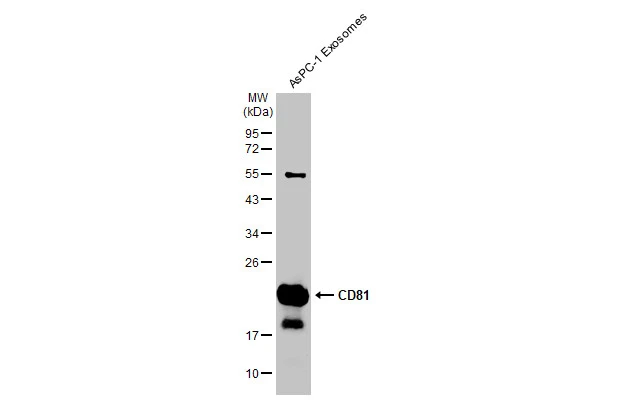
AsPC-1 exsosome extracts (5.5 μg) was separated by 12% SDS-PAGE, and the membrane was blotted with CD81 antibody (GTX135297) diluted at 1:500. The HRP-conjugated anti-rabbit IgG antibody (GTX213110-01) was used to detect the primary antibody.
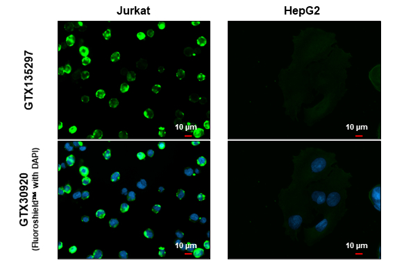
CD81 antibody detects CD81 protein by immunofluorescent analysis.Sample: Jurkat and HepG2 cells were fixed in 4% paraformaldehyde at RT for 15 min.Green: CD81 stained by CD81 antibody (GTX135297) diluted at 1:500.
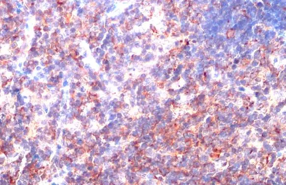
CD81 antibody detects CD81 protein at cell membrane by immunohistochemical analysis.Sample: Paraffin-embedded mouse thymus gland.CD81 stained by CD81 antibody (GTX135297) diluted at 1:500.Antigen Retrieval: Citrate buffer, pH 6.0, 15 min
-
HostRabbit
-
ClonalityPolyclonal
-
IsotypeIgG
-
ApplicationsWB ICC/IF IHC-P ELISA Sandwich ELISA
-
ReactivityHuman, Mouse