CDK4 antibody


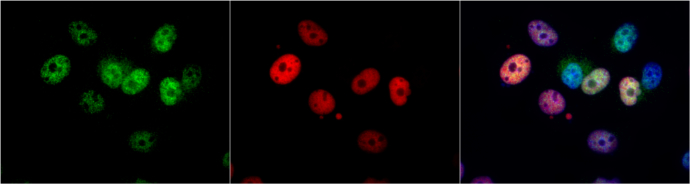
CDK4 antibody detects CDK4 protein at nucleus by immunofluorescent analysis.
Sample: MCF7 cells were fixed in 4% paraformaldehyde at RT for 15 min.
Green: CDK4 protein stained by CDK4 antibody (GTX102993) diluted at 1:1000.
Red: p21 Cip1, a nucleus marker, stained by p21 Cip1 antibody [GT1032] (GTX629543) diluted at 1:500.
Blue: Hoechst 33342 staining.
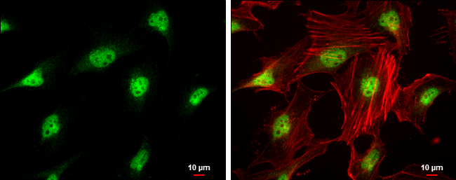
CDK4 antibody detects CDK4 protein at nucleus by immunofluorescent analysis.
Sample: HeLa cells were fixed in 4% paraformaldehyde at RT for 15 min.
Green: CDK4 protein stained by CDK4 antibody (GTX102993) diluted at 1:500.
Red: phalloidin, a cytoskeleton marker, diluted at 1:200.
Scale bar = 10 μm.
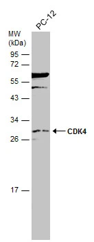
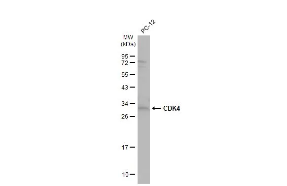
Whole cell extract (30 μg) was separated by 12% SDS-PAGE, and the membrane was blotted with CDK4 antibody (GTX102993) diluted at 1:500. The HRP-conjugated anti-rabbit IgG antibody (GTX213110-01) was used to detect the primary antibody.

Immunohistochemical analysis of paraffin-embedded human gastric cancer, using CDK4(GTX102993) antibody at 1:500 dilution.
Antigen Retrieval: Trilogy™ (EDTA based, pH 8.0) buffer, 15min
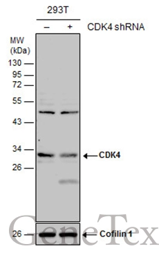
Non-transfected (–) and transfected (+) 293T whole cell extracts (30 μg) were separated by 12% SDS-PAGE, and the membrane was blotted with CDK4 antibody (GTX102993) diluted at 1:1000. The HRP-conjugated anti-rabbit IgG antibody (GTX213110-01) was used to detect the primary antibody.
Whole cell extract (30 μg) was separated by 12% SDS-PAGE, and the membrane was blotted with CDK4 antibody (GTX102993) diluted at 1:500. The HRP-conjugated anti-rabbit IgG antibody (GTX213110-01) was used to detect the primary antibody.
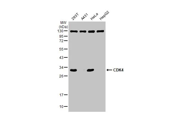
Various whole cell extracts (30 μg) were separated by 12% SDS-PAGE, and the membrane was blotted with CDK4 antibody (GTX102993) diluted at 1:1000. The HRP-conjugated anti-rabbit IgG antibody (GTX213110-01) was used to detect the primary antibody.
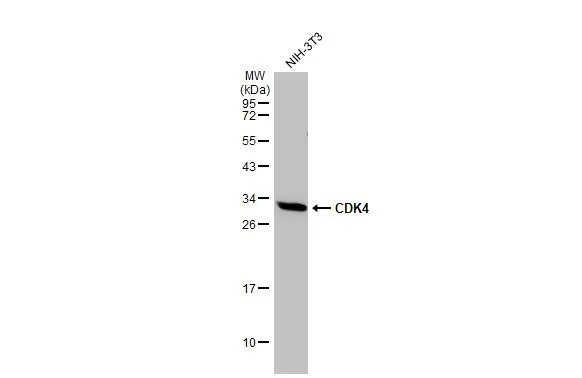
Whole cell extract (30 μg) was separated by 12% SDS-PAGE, and the membrane was blotted with CDK4 antibody (GTX102993) diluted at 1:500. The HRP-conjugated anti-rabbit IgG antibody (GTX213110-01) was used to detect the primary antibody.
-
HostRabbit
-
ClonalityPolyclonal
-
IsotypeIgG
-
ApplicationsWB ICC/IF IHC-P
-
ReactivityHuman, Mouse, Rat, Bovine