CDK9 antibody
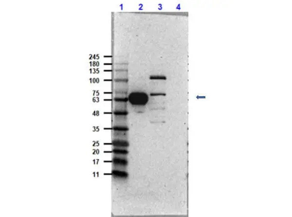
WB analysis of various samples using GTX26544 CDK9 antibody.
Lane 1 : Protein ladder
Lane 2 : Human kidney tissue lysate (5 μg)
Lane 3 : PC-3 whole cell lysate (20 μg)
Lane 4 : Null
Dilution : 1:1000
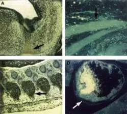
IHC-P analysis of various tissues using GTX26544 CDK9 antibody.
Panel A : Mouse brain
Panel B : Mouse dorsal root ganglia tissue section
Panel D : Mouse cardiac muscle tissue section
Dilution : 1:500
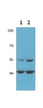
Genetex anti cdk9 antibody (GTX26544) was used for Western blot analysis of 1) PC3 and 2) DU145 prostate cancer cells (50ug per lane). Bands at the expected MW of 55 and 42 Kda were detected.

ICC staining of mouse tissue using anti-cdk9(PITALRE). The staining shows the location of mcdk9/PITALRE protein in developing mouse tissue. Arrows indicate areas of high expression. Panel A: Peroxidase-DAB immunostaining of mcdk9/PITALRE protein in the developing mouse brain in the differentiated region of the medulla oblongata just below the fourth ventricle. Similar staining is shown in Panel B in the dorsal root ganglia. Panel C: Fluorescein immunofluorescence of mcdk9IPITALRE in skeletal muscle. Similar staining is shown in Panel D in cardiac muscle. Sections from each specimen were cut at 5-7 μm, mounted on glass and dried overnight at 37ºC. All sections then were deparaffinized in xylene, rehydrated through a graded alcohol series and washed in phosphate-buffered saline (PBS). PBS was used for all subsequent washes and for antiserum dilution. Tissue sections were quenched sequentially in 0.5% hydrogen peroxide and blocked with diluted 10% normal goat anti-rabbit serum. Slides were incubated at 20º C for 1 h with rabbit anti-cdk9 (GTX26544)(1:500) dilution, washed, and then reacted with diluted goat anti-rabbit biotinylated antibody for 30 min. All the slides were then reacted with streptavidin-peroxidase conjugate for 30 min at 20º C. Diaminobenzidine was used as the final chromogen and hematoxylin was used as the nuclear counterstain. Negative controls for each tissue section were prepared by substituting the primary antiserum with pre-immune serum.
-
HostRabbit
-
ClonalityPolyclonal
-
IsotypeIgG
-
ApplicationsWB IHC-P IP ELISA
-
ReactivityHuman, Mouse