CIAPIN1 antibody
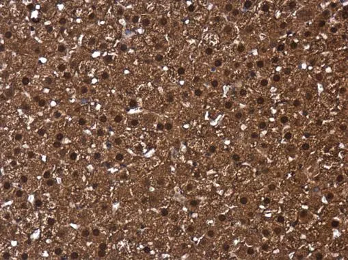
CIAPIN1 antibody detects CIAPIN1 protein at cytoplasm and nucleus in rat adrenal gland by immunohistochemical analysis.
Sample: Paraffin-embedded rat adrenal gland.
CIAPIN1 antibody (GTX111212) diluted at 1:500.
Antigen Retrieval: Citrate buffer, pH 6.0, 15 min
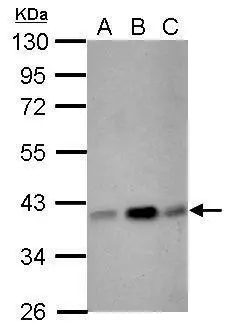
Sample (30 ug of whole cell lysate)
A: NIH-3T3
B: JC
C: BCL-1
10% SDS PAGE
GTX111212 diluted at 1:3000
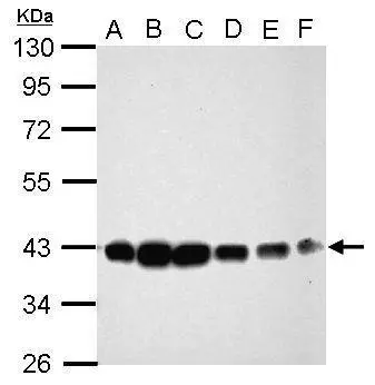
Sample (30 ug of whole cell lysate)
A: Jurkat
B: Raji
C: K562
D: THP-1
E: HL-60
F: NCI-H929
10% SDS PAGE
GTX111212 diluted at 1:1000
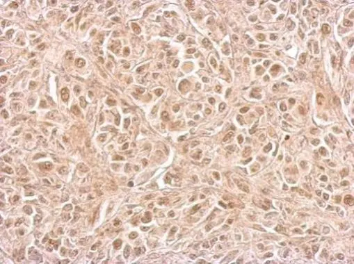
CIAPIN1 antibody detects CIAPIN1 protein at cytosol and nucleus on SkHep1 xenograft by immunohistochemical analysis.
Sample: Paraffin-embedded SkHep1 xenograft.
CIAPIN1 antibody (GTX111212) dilution: 1:500.
Antigen Retrieval: Trilogy™ (EDTA based, pH 8.0) buffer, 15min
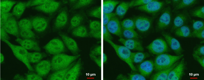
CIAPIN1 antibody detects CIAPIN1 protein at nucleus and cytoplasm by immunofluorescent analysis.
Sample: A375 cells were fixed in 4% paraformaldehyde at RT for 15 min.
Green: CIAPIN1 protein stained by CIAPIN1 antibody (GTX111212) diluted at 1:500.
Blue: Hoechst 33342 staining.
Scale bar = 10 μm.
-
HostRabbit
-
ClonalityPolyclonal
-
IsotypeIgG
-
ApplicationsWB ICC/IF IHC-P
-
ReactivityHuman, Mouse, Rat