COPD antibody

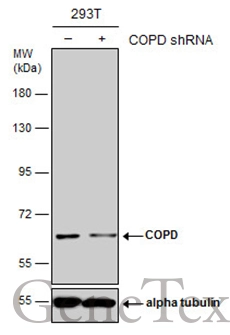
Non-transfected (–) and transfected (+) 293T whole cell extracts (30 μg) were separated by 7.5% SDS-PAGE, and the membrane was blotted with COPD antibody (GTX103252) diluted at 1:2000.
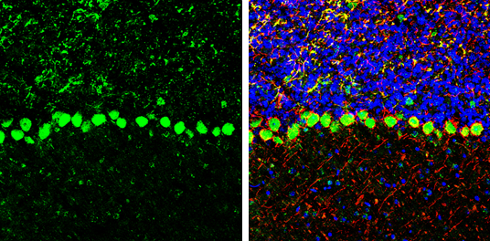
COPD antibody detects COPD protein by immunohistochemical analysis.Sample: Frozen-sectioned mouse mouse cerebellum.Green: COPD stained by COPD antibody (GTX103252) diluted at 1:250.Red: NF-H, stained by NF-H antibody [GT114] (GTX634289) diluted at 1:500.Blue: Fluoroshield with DAPI (GTX30920).
Antigen Retrieval: Citrate buffer, pH 6.0, 10 min
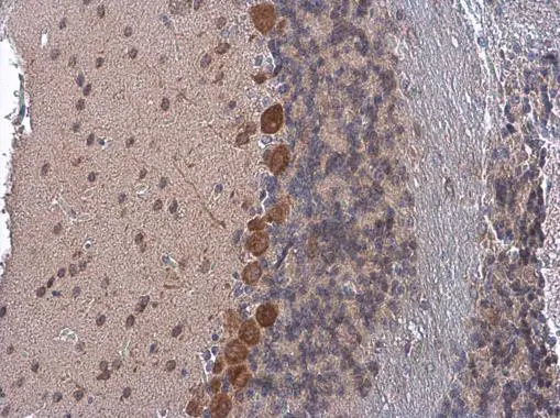
COPD antibody detects COPD protein at cytoplasm by immunohistochemical analysis.Sample: Paraffin-embedded mouse brain.COPD stained by COPD antibody (GTX103252) diluted at 1:500.
Antigen Retrieval: Citrate buffer, pH 6.0, 10 min
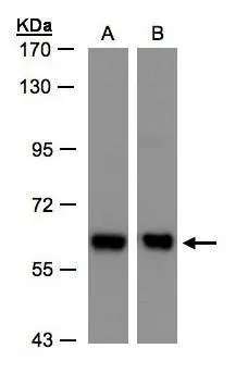
Sample(30 ug whole cell lysate)
A:H1299
B:Hep G2(GTX27900)
7.5% SDS PAGE
GTX103252 diluted at 1:500
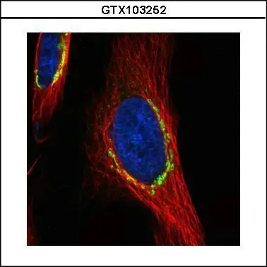
Confocal immunofluorescence analysis (Olympus FV10i) of paraformaldehyde-fixed HeLa, using COPD(GTX103252) antibody (green) at 1:500 dilution. Alpha-tubulin filaments were labeled with GTX11304 (red) at 1:2500.
-
HostRabbit
-
ClonalityPolyclonal
-
IsotypeIgG
-
ApplicationsWB ICC/IF IHC-P IHC-Fr
-
ReactivityHuman, Mouse