COX4 antibody


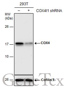
Non-transfected (–) and transfected (+) 293T whole cell extracts (30 μg) were separated by 15% SDS-PAGE, and the membrane was blotted with COX4 antibody (GTX114330) diluted at 1:3000. The HRP-conjugated anti-rabbit IgG antibody (GTX213110-01) was used to detect the primary antibody.
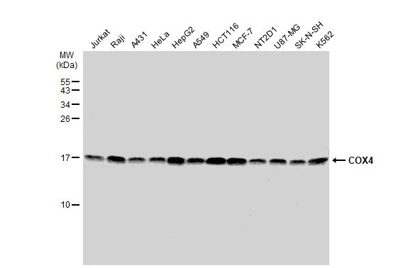
Various whole cell extracts (30 μg) were separated by 15% SDS-PAGE, and the membrane was blotted with COX4 antibody (GTX114330) diluted at 1:1000. The HRP-conjugated anti-rabbit IgG antibody (GTX213110-01) was used to detect the primary antibody.
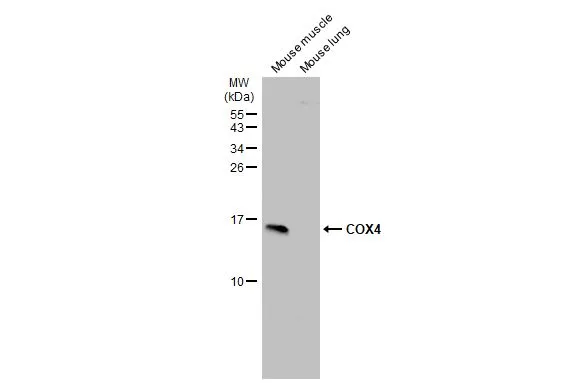
Various tissue extracts (50 μg) were separated by 15% SDS-PAGE, and the membrane was blotted with COX4 antibody (GTX114330) diluted at 1:1000. The HRP-conjugated anti-rabbit IgG antibody (GTX213110-01) was used to detect the primary antibody.
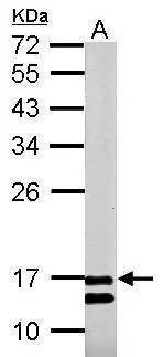
Sample (50 μg of whole cell lysate)
A: rat muscle
12% SDS PAGE
GTX114330 diluted at 1:500
The HRP-conjugated anti-rabbit IgG antibody (GTX213110-01) was used to detect the primary antibody.
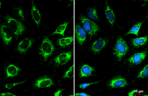
COX4 antibody detects COX4 protein at mitochondria by immunofluorescent analysis.Sample: HeLa cells were fixed in ice-cold MeOH for 5 min.Green: COX4 stained by COX4 antibody (GTX114330) diluted at 1:500.Blue: Hoechst 33342 staining.Scale bar= 10 μm.
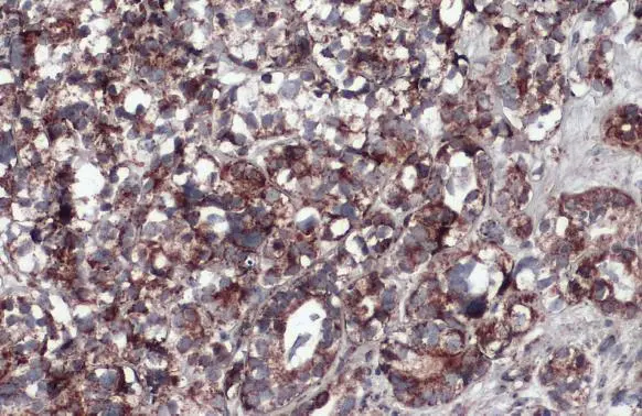
COX4 antibody detects COX4 protein at mitochondria by immunohistochemical analysis.Sample: Paraffin-embedded human endometrial carcinoma.COX4 stained by COX4 antibody (GTX114330) diluted at 1:500.Antigen Retrieval: Citrate buffer, pH 6.0, 15 min
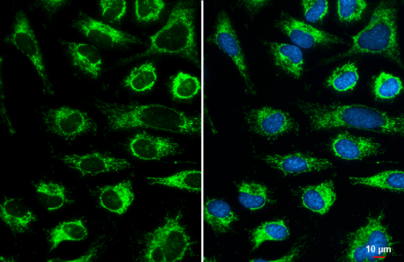
COX4 antibody detects COX4 protein at mitochondria by immunofluorescent analysis.Sample: HeLa cells were fixed in ice-cold MeOH for 5 min.Green: COX4 stained by COX4 antibody (GTX114330) diluted at 1:500.Blue: Hoechst 33342 staining.Scale bar= 10 μm.
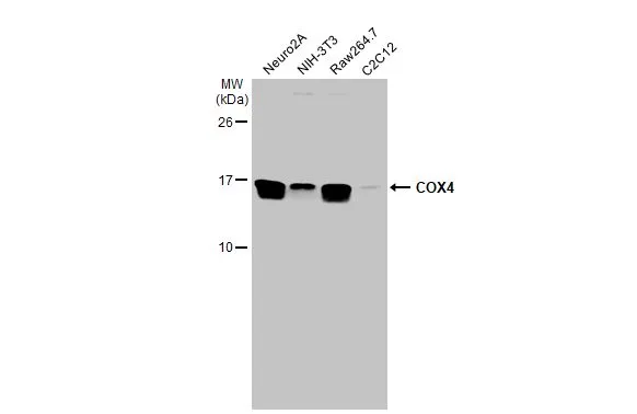
Various whole cell extracts (30 μg) were separated by 15% SDS-PAGE, and the membrane was blotted with COX4 antibody (GTX114330) diluted at 1:1000. The HRP-conjugated anti-rabbit IgG antibody (GTX213110-01) was used to detect the primary antibody.
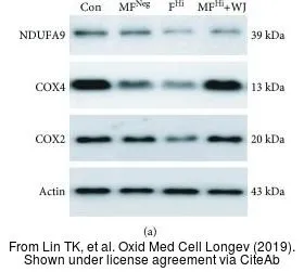
The data was published in the journal Oxid Med Cell Longev in 2019.PMID: 31249652
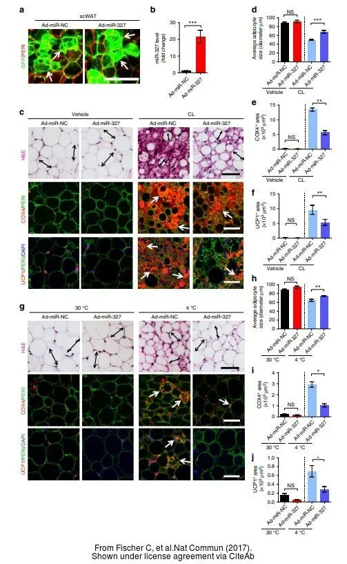
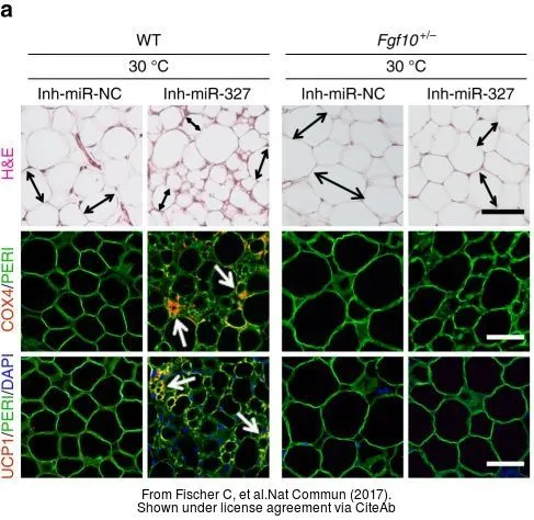
The data was published in the journal Nat Commun in 2017. PMID: 29233981
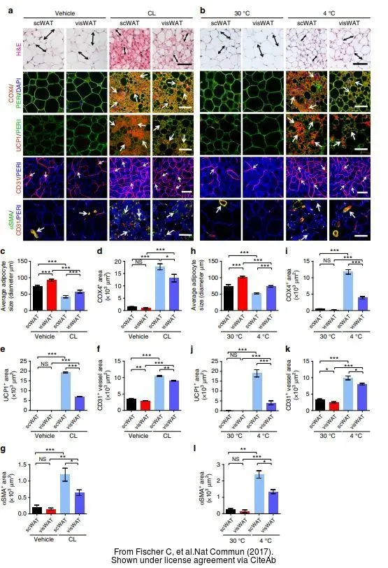
The data was published in the journal Nat Commun in 2017. PMID: 29233981
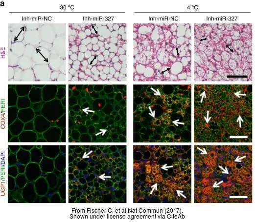
The data was published in the journal Nat Commun in 2017. PMID: 29233981
The data was published in the journal Nat Commun in 2017. PMID: 29233981
-
HostRabbit
-
ClonalityPolyclonal
-
IsotypeIgG
-
ApplicationsWB ICC/IF IHC-P IHC-Fr
-
ReactivityHuman, Mouse, Rat