Calponin 3 antibody


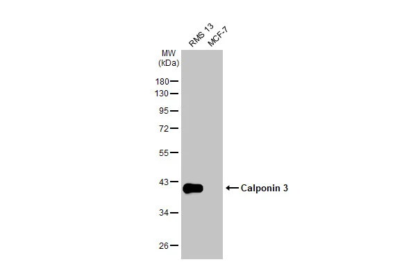
Various whole cell extracts (30 μg) were separated by 10% SDS-PAGE, and the membrane was blotted with Calponin 3 antibody (GTX106174) diluted at 1:1000. The HRP-conjugated anti-rabbit IgG antibody (GTX213110-01) was used to detect the primary antibody.
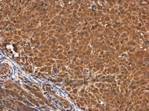
calponin 3 antibody detects calponin 3 protein at cytoplasm in rat ovary by immunohistochemical analysis.
Sample: Paraffin-embedded rat ovary.
calponin 3 antibody (GTX106174) diluted at 1:500.
Antigen Retrieval: Trilogy™ (EDTA based, pH 8.0) buffer, 15min
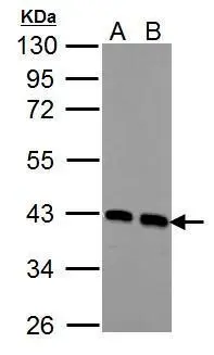
Sample (30 ug of whole cell lysate)
A: NIH-3T3
B: JC
10% SDS PAGE
GTX106174 diluted at 1:1000
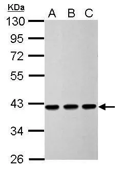
Sample (30 ug of whole cell lysate)
A: 293T
B: HeLa
C: HepG2
10% SDS PAGE
GTX106174 diluted at 1:5000
-
HostRabbit
-
ClonalityPolyclonal
-
IsotypeIgG
-
ApplicationsWB ICC/IF IHC-P IP
-
ReactivityHuman, Mouse, Rat