Cav1.2 antibody

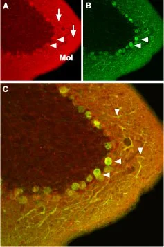
IHC-Fr analysis of mouse cerebellum tissue using GTX54754 Cav1.2 antibody.
Panel A : CaV1.2 (red) appears in Purkinje cells (horizontal arrows) and is distributed diffusely in the molecular layer (Mol) including in Purkinje dendrites (vertical arrows).
Panel B : Staining of Purkinje nerve cells with mouse anti-Calbindin 28K (green) demonstrates the location of dendrites in the molecular layer.
Panel C : Merged image of panels A and B.
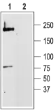
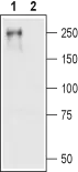
WB analysis of rat brain membrane lysate using GTX54754 Cav1.2 antibody preincubated with or without immunogen peptide.
Dilution : 1:200
-
HostRabbit
-
ClonalityPolyclonal
-
IsotypeIgG
-
ApplicationsWB ICC/IF IHC-Fr FCM IP
-
ReactivityHuman, Mouse, Rat