Cav1.3 antibody
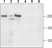
WB analysis of rat brain (lanes 1 and 4), mouse brain (lanes 2 and 5) and C6 (lanes 3 and 6) lysates using GTX16633 Cav1.3 antibody preincubated with or without immunogen peptide.
Dilution : 1:200
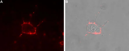
Live cell imaging analysis of intact living PC12 cells using GTX16633 Cav1.3 antibody.
Panel A : Primary antibody
Panel B : Merge of panel A with the live view of the cell
Dilution : 1:50
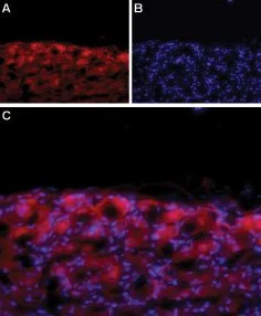
IHC-Fr analysis of rat DRG tissue using GTX16633 Cav1.3 antibody.
Panel A : CaV1.3 labeling (red) appears in the cell bodies of the DRG.
Panel B : Nuclear staining using DAPI as the counterstain.
Panel C : Merged image of A and B.
-
HostRabbit
-
ClonalityPolyclonal
-
IsotypeIgG
-
ApplicationsWB ICC/IF IHC-Fr LCI
-
ReactivityHuman, Mouse, Rat