Collagen I antibody (Biotin)
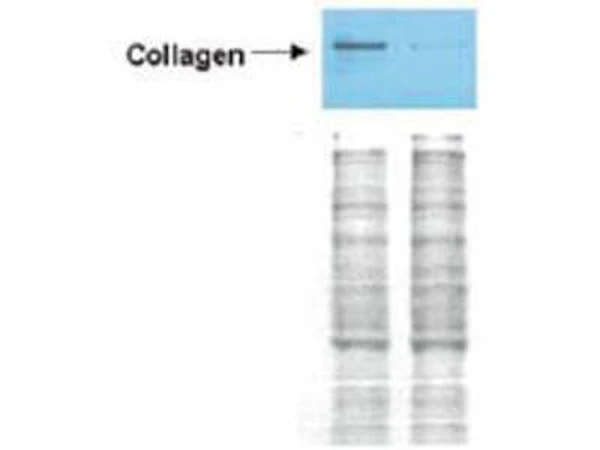
WB analysis of Wistar rat hepatic stellate whole cell lysate using GTX26577 Collagen I antibody (Biotin).
Left lane : GFP-transduced
Right lane : PPARg-transduced
Loading : 100 μg
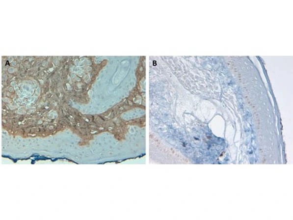
IHC-P analysis of human skin tissue at pH 6 using GTX26577 Collagen I antibody (Biotin).
Panel A : Positive control
Panel B : Negative control
Dilution : 10 μgm/mL
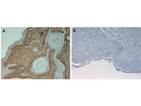
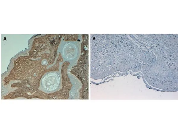
IHC-P analysis of human skin tissue at pH 9 using GTX26577 Collagen I antibody (Biotin).
Panel A : Positive control
Panel B : Negative control
Dilution : 10 μg/mL

Flow Cytometry of Rabbit Anti-Collagen 1 Antibody. Cells: primary adult human dermal fibroblast cells. Stimulation: none. Primary antibody: Biotin-Conjugated Collagen 1 antibody at 5μg/mL for 45 min at 4ºC. Secondary antibody: Rabbit Streptavidin, R-PE antibody at 1:500 for 15 min at RT.
Immunohistochemistry of Rabbit Anti-Collagen Type I Antibody (GTX26577). Tissue: Human Skin at pH6. Fixation: formalin fixed paraffin embedded. Antigen retrieval: not required. Primary antibody: Collagen Type I antibody at 10 μg/mL for 1 h at RT. Secondary antibody: Peroxidase rabbit secondary antibody at 1:10,000 for 45 min at RT. Localization: Collagen Type I is secreted in the extracellular matrix. Staining: Collagen Type I as precipitated brown signal (A) with hematoxylin purple nuclear counterstain. With corresponding negative conrol (B)

Immunohistochemistry of Rabbit Anti-Collagen Type I Antibody (GTX26577). Tissue: Human Skin at pH9. Fixation: formalin fixed paraffin embedded. Antigen retrieval: not required. Primary antibody: Collagen Type I antibody at 10 μg/mL for 1 h at RT. Secondary antibody: Peroxidase rabbit secondary antibody at 1:10,000 for 45 min at RT. Localization: Collagen Type I is secreted in the extracellular matrix. Staining: Collagen Type I as precipitated brown signal (A) with hematoxylin purple nuclear counterstain. With corresponding negative control (B)
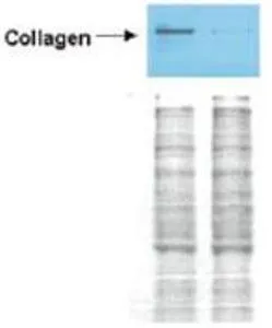
Western Blot of Rabbit anti-Collagen I antibody (GTX26577). Lane 1: Wistar rat hepatic stellate cells (HSC) in control (GFP-transduced). Lane 2: PPARg-transduced cell lysates. Load: 100 μg per lane. Protein staining shown below each blot depicts equal protein loading. Primary antibody: anti-Collagen I antibody at 0.2–2 μg/10 ml for overnight at 4ºC. Secondary antibody: horseradish peroxidase-conjugated rabbit secondary antibody at 1 μg/10 ml for overnight at 4ºC. Block: TBS with 5% Non-fat milk. Predicted/Observed size: 138.9 kDa for Collagen I. Other band(s): none.
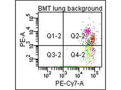
Flow Cytometry of Anti-Collagen Type I Biotin Conjugated Antibody. Cells: mouse lung. Stimulation: none. Primary antibody: biotin conjugated anti-collagen type I antibody. Secondary antibody: PE-conjugated CD45 and PE-conjugated anti-collagen type I secondary antibody.
-
HostRabbit
-
ClonalityPolyclonal
-
IsotypeIgG
-
ApplicationsWB ICC/IF IHC-P FCM Dot Multiplexing
-
ReactivityHuman, Mouse, Rat