Coxsackie Adenovirus Receptor antibody
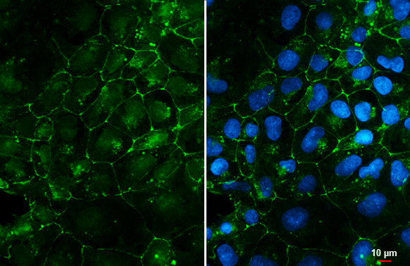
Coxsackie Adenovirus Receptor antibody detects Coxsackie Adenovirus Receptor protein at cell membrane by immunofluorescent analysis.Sample: NT2D1 cells were fixed in 4% paraformaldehyde at RT for 15 min.Green: Coxsackie Adenovirus Receptor stained by Coxsackie Adenovirus Receptor antibody (GTX118382) diluted at 1:500.Blue: Fluoroshield with DAPI (GTX30920).Scale bar= 10 μm.
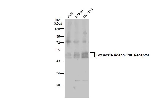
Various whole cell extracts (30 μg) were separated by 10% SDS-PAGE, and the membrane was blotted with Coxsackie Adenovirus Receptor antibody (GTX118382) diluted at 1:1000. The HRP-conjugated anti-rabbit IgG antibody (GTX213110-01) was used to detect the primary antibody.
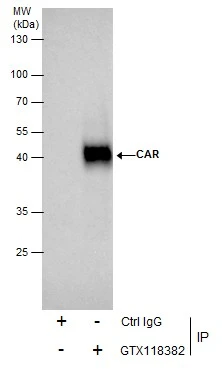
Immunoprecipitation of CAR protein from HeLa whole cell extracts using 5 μg of CAR antibody (GTX118382).
Western blot analysis was performed using CAR antibody (GTX118382).
EasyBlot anti-Rabbit IgG (GTX221666-01) was used as a secondary reagent.
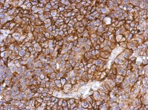
Immunohistochemical analysis of paraffin-embedded PC13 xenograft, using CAR(GTX118382) antibody at 1:500 dilution.
Antigen Retrieval: Trilogy™ (EDTA based, pH 8.0) buffer, 15min
-
HostRabbit
-
ClonalityPolyclonal
-
IsotypeIgG
-
ApplicationsWB ICC/IF IHC-P IP
-
ReactivityHuman, Rat