Cytochrome C antibody


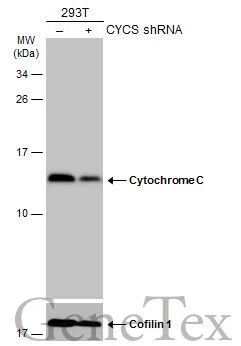
Non-transfected (–) and transfected (+) 293T whole cell extracts (30 μg) were separated by 15% SDS-PAGE, and the membrane was blotted with Cytochrome C antibody (GTX108585) diluted at 1:1000. The HRP-conjugated anti-rabbit IgG antibody (GTX213110-01) was used to detect the primary antibody.
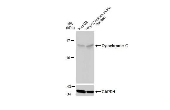
HepG2 and mitochondria extracts (30 μg) were separated by SDS-PAGE, and the membrane was blotted with Cytochrome C antibody (GTX108585) diluted at 1:2000. The HRP-conjugated anti-rabbit IgG antibody (GTX213110-01) was used to detect the primary antibody, and the signal was developed with Trident pico Western HRP Substrate.
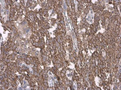
Cytochrome C antibody detects Cytochrome C protein at cytoplasm by immunohistochemical analysis.Sample: Paraffin-embedded human endometrial carcinoma.Cytochrome C stained by Cytochrome C antibody (GTX108585) diluted at 1:500.
Antigen Retrieval: Citrate buffer, pH 6.0, 15 min
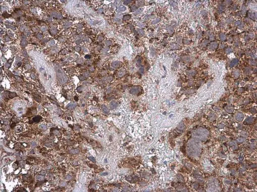
Cytochrome C antibody detects Cytochrome C protein at cytoplasm by immunohistochemical analysis.Sample: Paraffin-embedded human cervical carcinoma.Cytochrome C stained by Cytochrome C antibody (GTX108585) diluted at 1:500.
Antigen Retrieval: Citrate buffer, pH 6.0, 15 min
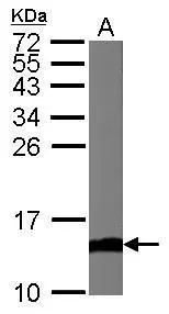
Sample (50 μg of whole cell lysate)
A: Mouse brain
15% SDS PAGE
GTX108585 diluted at 1:1000
The HRP-conjugated anti-rabbit IgG antibody (GTX213110-01) was used to detect the primary antibody.
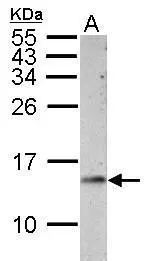
Sample (30 μg of whole cell lysate)
A: PC-12
15% SDS PAGE
GTX108585 diluted at 1:1000
The HRP-conjugated anti-rabbit IgG antibody (GTX213110-01) was used to detect the primary antibody.
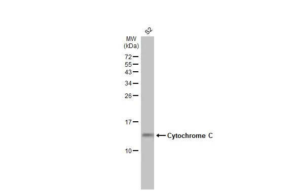
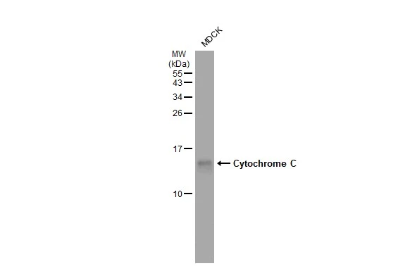
Whole cell extract (30 μg) was separated by 15% SDS-PAGE, and the membrane was blotted with Cytochrome C antibody (GTX108585) diluted at 1:1000. The HRP-conjugated anti-rabbit IgG antibody (GTX213110-01) was used to detect the primary antibody, and the signal was developed with Trident ECL plus-Enhanced.
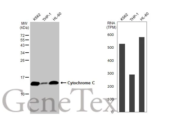
Various whole cell extracts (30 μg) were separated by 15% SDS-PAGE, and the membrane was blotted with Cytochrome C antibody (GTX108585) diluted at 1:1000. The HRP-conjugated anti-rabbit IgG antibody (GTX213110-01) was used to detect the primary antibody, and the signal was developed with Trident ECL plus-Enhanced. Corresponding RNA expression data for the same cell lines are based on Human Protein Atlas program.
Whole cell extract (30 μg) was separated by 15% SDS-PAGE, and the membrane was blotted with Cytochrome C antibody (GTX108585) diluted at 1:1000. The HRP-conjugated anti-rabbit IgG antibody (GTX213110-01) was used to detect the primary antibody.
-
HostRabbit
-
ClonalityPolyclonal
-
IsotypeIgG
-
ApplicationsWB ICC/IF IHC-P IHC-Fr
-
ReactivityHuman, Mouse, Rat, Drosophila, Dog