Cytokeratin 8 antibody

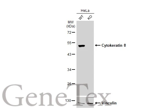
Wild-type (WT) and Cytokeratin 8 knockout (KO) HeLa cell extracts (30 μg) were separated by 10% SDS-PAGE, and the membrane was blotted with Cytokeratin 8 antibody (GTX110311) diluted at 1:20000. The HRP-conjugated anti-rabbit IgG antibody (GTX213110-01) was used to detect the primary antibody.
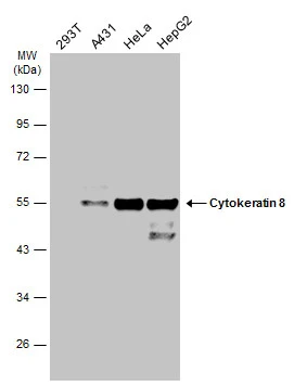
Various whole cell extracts (30 μg) were separated by 10% SDS-PAGE, and the membrane was blotted with Cytokeratin 8 antibody (GTX110311) diluted at 1:10000. The HRP-conjugated anti-rabbit IgG antibody (GTX213110-01) was used to detect the primary antibody.
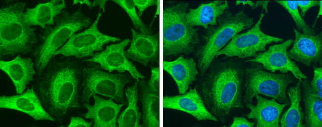
Cytokeratin 8 antibody detects Cytokeratin 8 protein at cytoskeleton by immunofluorescent analysis.Sample: HeLa cells were fixed in 4% paraformaldehyde at RT for 15 min.Green: Cytokeratin 8 stained by Cytokeratin 8 antibody (GTX110311) diluted at 1:500.Blue: Hoechst 33342 staining.
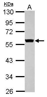
Sample (30 μg of whole cell lysate)
A: JC
10% SDS PAGE
GTX110311 diluted at 1:10000
The HRP-conjugated anti-rabbit IgG antibody (GTX213110-01) was used to detect the primary antibody.
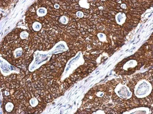
Immunohistochemical analysis of paraffin-embedded Gastric ca, using Cytokeratin 8(GTX110311) antibody at 1:500 dilution.
Antigen Retrieval: Trilogy™ (EDTA based, pH 8.0) buffer, 15min
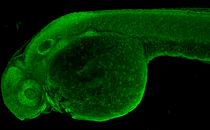
Cytokeratin 8 antibody detects Cytokeratin 8 protein on whole mount zebrafish by immunohistochemical analysis.Sample: Paraformaldehyde-fixed 2 days-post-fertilization zebrafish embryo.Green: Cytokeratin 8 stained by Cytokeratin 8 antibody (GTX110311) diluted at 1:100.
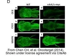
The data was published in the journal Oncotarget in 2014. PMID: 25051368
-
HostRabbit
-
ClonalityPolyclonal
-
IsotypeIgG
-
ApplicationsWB ICC/IF IHC-P IHC-Wm
-
ReactivityHuman, Mouse, Zebrafish