DBT antibody
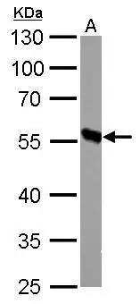
DBT antibody detects DBT protein by western blot analysis.
A. 50 μg mouse liver lysate/extract
10% SDS-PAGE
DBT antibody (GTX113412) dilution: 1:1000
The HRP-conjugated anti-rabbit IgG antibody (GTX213110-01) was used to detect the primary antibody.
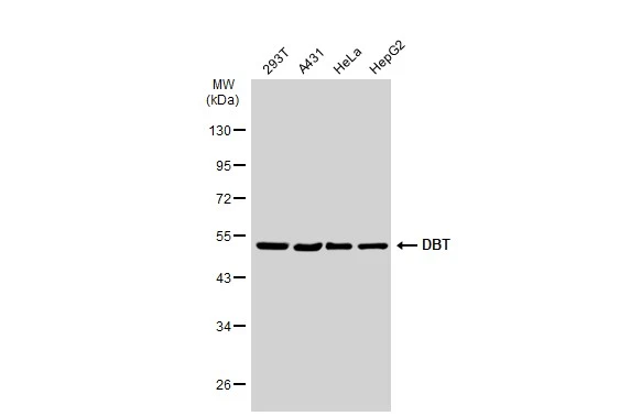
Various whole cell extracts (30 μg) were separated by 10% SDS-PAGE, and the membrane was blotted with DBT antibody (GTX113412) diluted at 1:1000. The HRP-conjugated anti-rabbit IgG antibody (GTX213110-01) was used to detect the primary antibody.
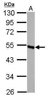
DBT antibody detects DBT protein by western blot analysis.
A. 50 μg rat liver lysate/extract
10% SDS-PAGE
DBT antibody (GTX113412) dilution: 1:1000
The HRP-conjugated anti-rabbit IgG antibody (GTX213110-01) was used to detect the primary antibody.
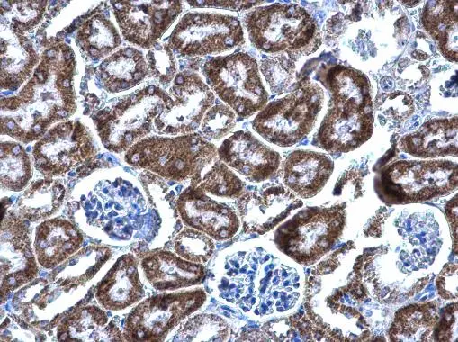
DBT antibody detects DBT protein at mitochondria on mouse kidney by immunohistochemical analysis.
Sample: Paraffin-embedded mouse kidney.
DBT antibody (GTX113412) diluted at 1:500.
Antigen Retrieval: Trilogy™ (EDTA based, pH 8.0) buffer, 15min
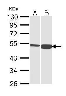
Sample (30 μg of whole cell lysate)
A: H1299
B: HepG2 (GTX27900)
10% SDS PAGE
GTX113412 diluted at 1:1000
The HRP-conjugated anti-rabbit IgG antibody (GTX213110-01) was used to detect the primary antibody.
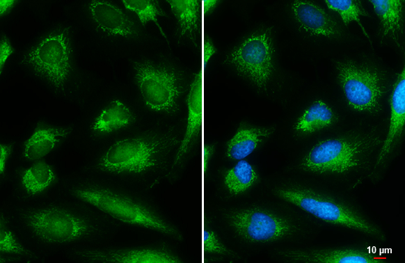
DBT antibody detects DBT protein at mitochondria by immunofluorescent analysis.Sample: HeLa cells were fixed in 4% paraformaldehyde at RT for 15 min.Green: DBT stained by DBT antibody (GTX113412) diluted at 1:500.Blue: Hoechst 33342 staining.Scale bar= 10 μm.
-
HostRabbit
-
ClonalityPolyclonal
-
IsotypeIgG
-
ApplicationsWB ICC/IF IHC-P
-
ReactivityHuman, Mouse, Rat