DDX1 antibody
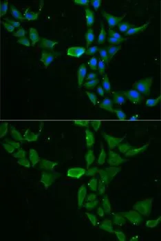
ICC/IF analysis of MCF7 cells using GTX33149 DDX1 antibody.
Blue : DAPI
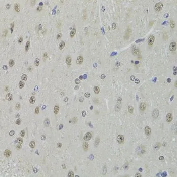
IHC-P analysis of mouse brain tissue using GTX33149 DDX1 antibody.
Dilution : 1:100
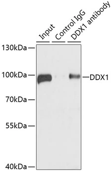
IP analysis of 293T cell lysate using GTX33149 DDX1 antibody.
Antibody amount : 3μg / 100μg lysate
Dilution : 1:1000
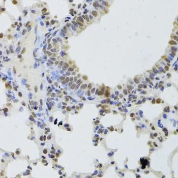
IHC-P analysis of mouse lung tissue using GTX33149 DDX1 antibody.
Dilution : 1:100
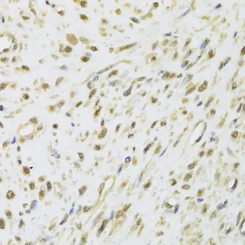
IHC-P analysis of human leiomyoma of uterus tissue using GTX33149 DDX1 antibody.
Dilution : 1:100
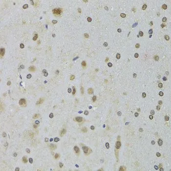
IHC-P analysis of rat brain tissue using GTX33149 DDX1 antibody.
Dilution : 1:100
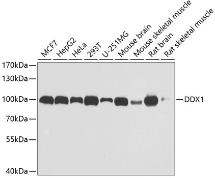
WB analysis of various sample lysates using GTX33149 DDX1 antibody.
Dilution : 1:1000
Loading : 25μg per lane
-
HostRabbit
-
ClonalityPolyclonal
-
IsotypeIgG
-
ApplicationsWB ICC/IF IHC-P IP
-
ReactivityHuman, Mouse, Rat