DIS3 antibody
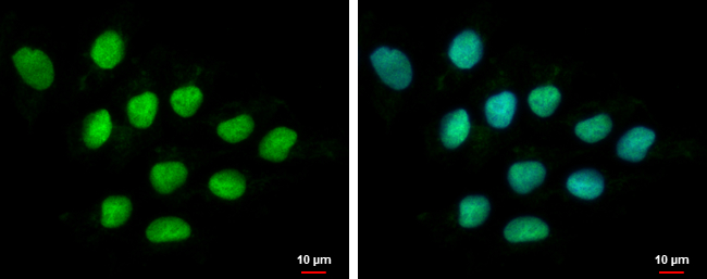
DIS3 antibody detects DIS3 protein at nucleus by immunofluorescent analysis.
Sample: NT2D1 cells were fixed in 4% paraformaldehyde at RT for 15 min.
Green: DIS3 protein stained by DIS3 antibody (GTX115645) diluted at 1:500.
Blue: Hoechst 33342 staining.
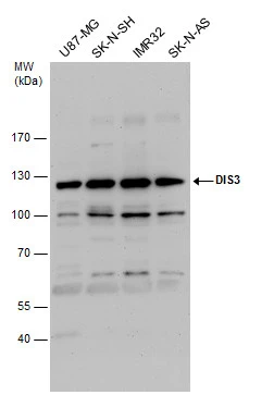
DIS3 antibody detects DIS3 protein by western blot analysis. Various whole cell extracts (30 μg) were separated by 7.5% SDS-PAGE, and the membrane was blotted with DIS3 antibody (GTX115645) diluted by 1:1000.
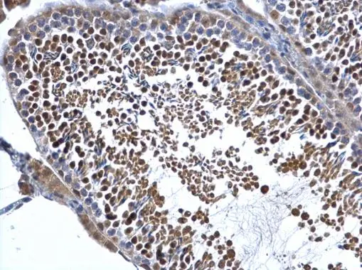
DIS3 antibody detects DIS3 protein at cytosol and nucleus on mouse testis by immunohistochemical analysis.
Sample: Paraffin-embedded mouse testis.
DIS3 antibody (GTX115645) dilution: 1:500.
Antigen Retrieval: Trilogy™ (EDTA based, pH 8.0) buffer, 15min
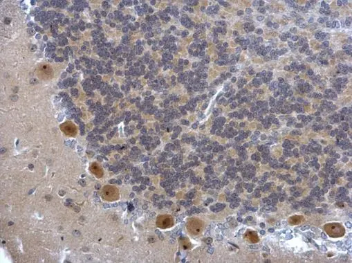
DIS3 antibody detects DIS3 protein at cytosol and nucleus on rat hind brain by immunohistochemical analysis.
Sample: Paraffin-embedded rat hind brain.
DIS3 antibody (GTX115645) dilution: 1:500.
Antigen Retrieval: Trilogy™ (EDTA based, pH 8.0) buffer, 15min
-
HostRabbit
-
ClonalityPolyclonal
-
IsotypeIgG
-
ApplicationsWB ICC/IF IHC-P
-
ReactivityHuman, Mouse, Rat