DLGAP1 antibody
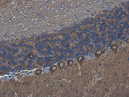
DLGAP1 antibody detects DLGAP1 protein at cytoplasm in mouse brain by immunohistochemical analysis.
Sample: Paraffin-embedded mouse brain.
DLGAP1 antibody (GTX133264) diluted at 1:500.
Antigen Retrieval: Citrate buffer, pH 6.0, 15 min
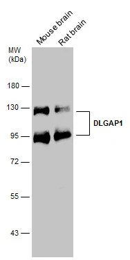
The observed M.W. is based on the publication: PMID: 21041448
Various tissue extracts (50 μg) were separated by 7.5% SDS-PAGE, and the membrane was blotted with DLGAP1 antibody (GTX133264) diluted at 1:3000.
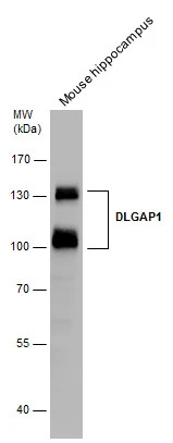
Mouse tissue extract (50 μg) was separated by 7.5% SDS-PAGE, and the membrane was blotted with DLGAP1 antibody (GTX133264) diluted at 1:3000.
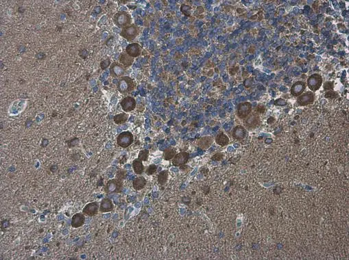
DLGAP1 antibody detects DLGAP1 protein at cytoplasm in rat brain by immunohistochemical analysis.
Sample: Paraffin-embedded rat brain.
DLGAP1 antibody (GTX133264) diluted at 1:500.
Antigen Retrieval: Citrate buffer, pH 6.0, 15 min
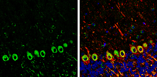
DLGAP1 antibody detects DLGAP1 Protein expression by immunohistochemical analysis.
Sample: Frozen-sectioned adult mouse cerebellum.
Green: DLGAP1 stained by DLGAP1 antibody (GTX133264) diluted at 1:250.
Red: NF-H, stained by NF-H antibody [GT114] (GTX634289) diluted at 1:500.
Blue: Fluoroshield with DAPI (GTX30920).
Antigen Retrieval: Citrate buffer, pH 6.0, 10 min
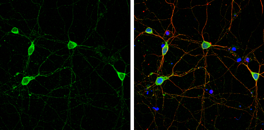
DLGAP1 antibody detects DLGAP1 protein by immunofluorescent analysis.Sample: DIV9 rat E18 primary cortical neuron cells were fixed in 4% paraformaldehyde at RT for 15 min.Green: DLGAP1 stained by DLGAP1 antibody (GTX133264) diluted at 1:500.Red: beta Tubulin 3/ Tuj1, stained by beta Tubulin 3/ Tuj1 antibody [GT1338] (GTX631831) diluted at 1:500.Blue: Fluoroshield with DAPI (GTX30920).
-
HostRabbit
-
ClonalityPolyclonal
-
IsotypeIgG
-
ApplicationsWB ICC/IF IHC-P IHC-Fr
-
ReactivityMouse, Rat