DR3 antibody
Cat. No. GTX105713
Cat. No. GTX105713
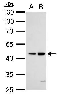
GTX105713 WB Image
DR3 antibody detects TNFRSF25 protein by Western blot analysis.
A. 30 μg Raji whole cell lysate/extract
B. 30 μg K562 whole cell lysate/extract
10 % SDS-PAGE
DR3 antibody (GTX105713) dilution: 1:1000
1 / 2
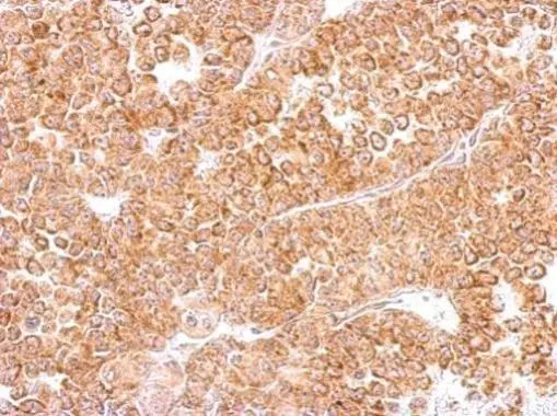
GTX105713 IHC-P Image
DR3 antibody detects TNFRSF25 protein at cytosol on AGS xenograft by immunohistochemical analysis.
Sample: Paraffin-embedded AGS xenograft.
DR3 antibody (GTX105713) dilution: 1:500.
Antigen Retrieval: Trilogy™ (EDTA based, pH 8.0) buffer, 15min
2 / 2
-
HostRabbit
-
ClonalityPolyclonal
-
IsotypeIgG
-
ApplicationsWB IHC-P
-
ReactivityHuman