DRP1 antibody
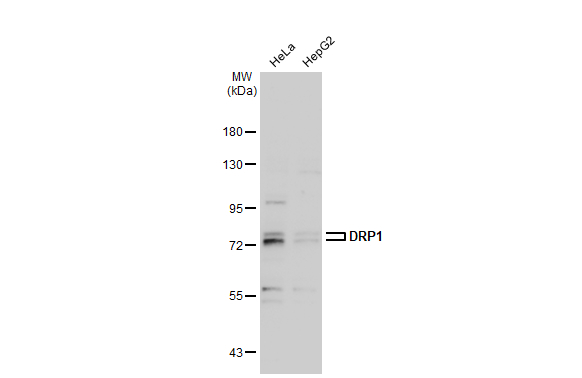
Various whole cell extracts (30 μg) were separated by 7.5% SDS-PAGE, and the membrane was blotted with DRP1 antibody (GTX135364) diluted at 1:2000. The HRP-conjugated anti-rabbit IgG antibody (GTX213110-01) was used to detect the primary antibody.
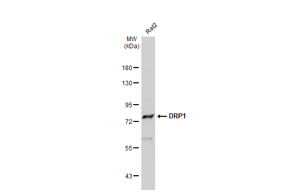
Mouse tissue extract (30 μg) was separated by 7.5% SDS-PAGE, and the membrane was blotted with DRP1 antibody (GTX135364) diluted at 1:2000. The HRP-conjugated anti-rabbit IgG antibody (GTX213110-01) was used to detect the primary antibody.
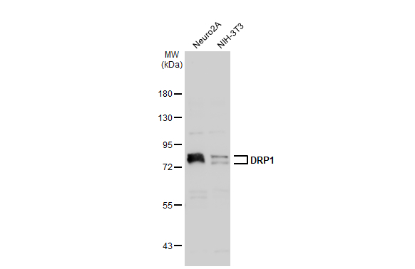
Various whole cell extracts (30 μg) were separated by 7.5% SDS-PAGE, and the membrane was blotted with DRP1 antibody (GTX135364) diluted at 1:2000. The HRP-conjugated anti-rabbit IgG antibody (GTX213110-01) was used to detect the primary antibody.
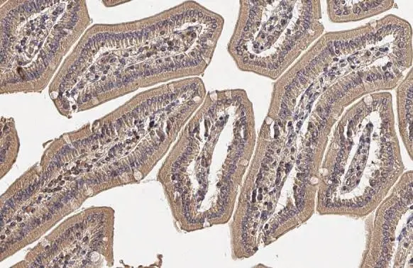
DRP1 antibody detects DRP1 protein by immunohistochemical analysis.Sample: Paraffin-embedded mouse duodenum.DRP1 stained by DRP1 antibody (GTX135364) diluted at 1:500.Antigen Retrieval: Citrate buffer, pH 6.0, 15 min

DRP1 antibody detects DRP1 protein by immunohistochemical analysis.Sample: Paraffin-embedded rat stomach.DRP1 stained by DRP1 antibody (GTX135364) diluted at 1:500.Antigen Retrieval: Citrate buffer, pH 6.0, 15 min
-
HostRabbit
-
ClonalityPolyclonal
-
IsotypeIgG
-
ApplicationsWB IHC-P
-
ReactivityHuman, Mouse, Rat