E-Cadherin antibody


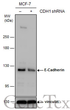
Non-transfected (–) and transfected (+) MCF-7 whole cell extracts (30 μg) were separated by 5% SDS-PAGE, and the membrane was blotted with E-Cadherin antibody (GTX100443) diluted at 1:7000. The HRP-conjugated anti-rabbit IgG antibody (GTX213110-01) was used to detect the primary antibody.
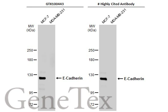
Various whole cell extracts (30 μg) were separated by 5% SDS-PAGE, and the membranes were blotted with E-Cadherin antibody (GTX100443) diluted at 1:3000 and competitor's antibody diluted at 1:3000. The HRP-conjugated anti-rabbit IgG antibody (GTX213110-01) was used to detect the primary antibody.
*The competitor is not affiliated with GeneTex and does not endorse this product.
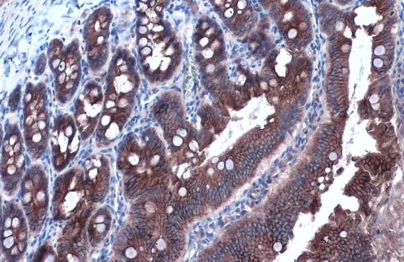
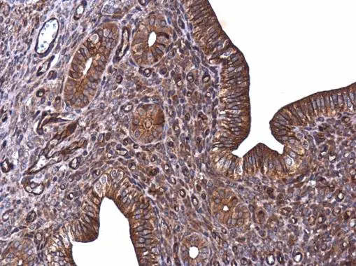
E-Cadherin antibody detects E-Cadherin protein at cell membrane and cytoplasm by immunohistochemical analysis.Sample: Paraffin-embedded rat duodenum.E-Cadherin stained by E-Cadherin antibody (GTX100443) diluted at 1:1000.Antigen Retrieval: Citrate buffer, pH 6.0, 15 min
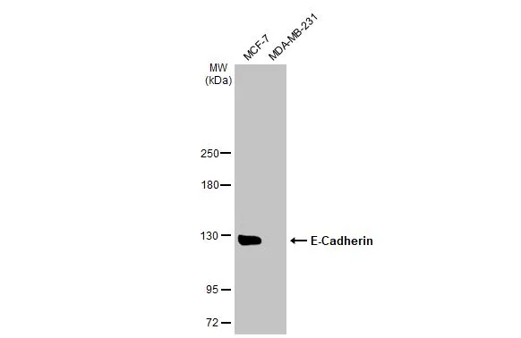
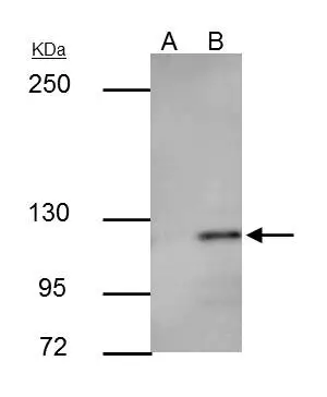
Various whole cell extracts (30 μg) were separated by 5% SDS-PAGE, and the membrane was blotted with E-Cadherin antibody (GTX100443) diluted at 1:2000. The HRP-conjugated anti-rabbit IgG antibody (GTX213110-01) was used to detect the primary antibody.
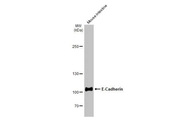
Mouse tissue extract (50 μg) was separated by 5% SDS-PAGE, and the membrane was blotted with E-Cadherin antibody (GTX100443) diluted at 1:2000. The HRP-conjugated anti-rabbit IgG antibody (GTX213110-01) was used to detect the primary antibody.
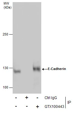
Immunoprecipitation of E-Cadherin protein from MCF-7 whole cell extracts using 5 μg of E-Cadherin antibody (GTX100443).
Western blot analysis was performed using E-Cadherin antibody (GTX100443).
EasyBlot anti-Rabbit IgG (GTX221666-01) was used as a secondary reagent.
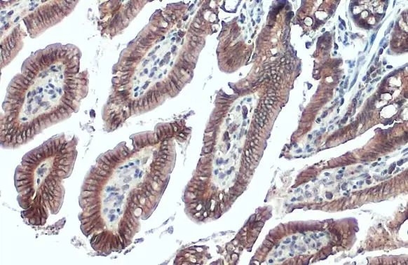
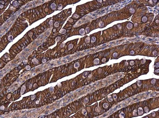
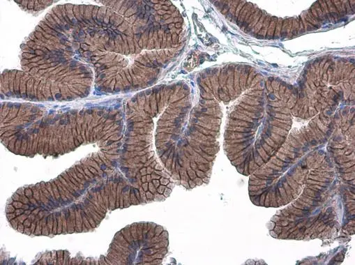
E-Cadherin antibody detects E-Cadherin protein at cell membrane and cytoplasm by immunohistochemical analysis.Sample: Paraffin-embedded mouse intestine.E-Cadherin stained by E-Cadherin antibody (GTX100443) diluted at 1:500.Antigen Retrieval: Citrate buffer, pH 6.0, 15 min
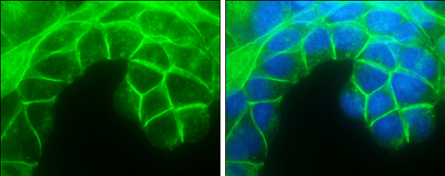
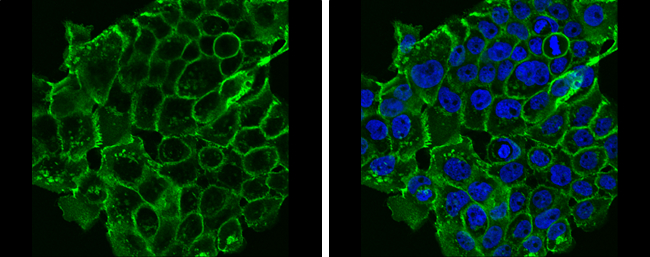
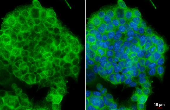
E-Cadherin antibody detects E-Cadherin protein at cell membrane by immunofluorescent analysis.
Sample: MCF7 cells were fixed in 4% paraformaldehyde at RT for 15 min.
Green: E-Cadherin protein stained by E-Cadherin antibody (GTX100443) diluted at 1:500.
Blue: Hoechst 33342 staining.
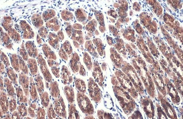
E-Cadherin antibody detects E-Cadherin protein at cell membrane and cytoplasm by immunohistochemical analysis.Sample: Paraffin-embedded mouse stomach.E-Cadherin stained by E-Cadherin antibody (GTX100443) diluted at 1:500.Antigen Retrieval: Citrate buffer, pH 6.0, 15 min
E-Cadherin antibody detects E-Cadherin protein at cell membrane in mouse cervix by immunohistochemical analysis.
Sample: Paraffin-embedded mouse cervix.
E-Cadherin antibody (GTX100443) diluted at 1:500.
Antigen Retrieval: Citrate buffer, pH 6.0, 15 min
E-Cadherin antibody detects E-Cadherin protein at cell membrane in rat intestine by immunohistochemical analysis.
Sample: Paraffin-embedded rat intestine.
E-Cadherin antibody (GTX100443) diluted at 1:500.
Antigen Retrieval: Citrate buffer, pH 6.0, 15 min
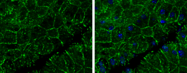
E-Cadherin antibody detects E-Cadherin protein at cell membrane by immunofluorescent analysis.
Sample: A431 cells were fixed in 4% paraformaldehyde at RT for 15 min.
Green: E-Cadherin protein stained by E-Cadherin antibody (GTX100443) diluted at 1:500.
Blue: Hoechst 33342 staining.
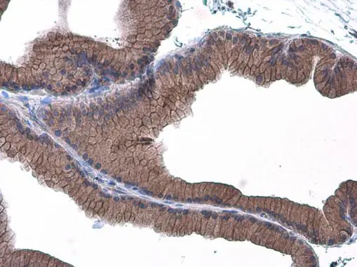
E-Cadherin antibody detects E-Cadherin protein at cell membrane in mouse pancreas by immunohistochemical analysis.
Sample: Paraffin-embedded mouse pancreas.
E-Cadherin antibody (GTX100443) diluted at 1:400.
Antigen Retrieval: Citrate buffer, pH 6.0, 15 min
E-Cadherin antibody detects E-Cadherin protein at cell membrane in rat prostate by immunohistochemical analysis.
Sample: Paraffin-embedded rat prostate.
E-Cadherin antibody (GTX100443) diluted at 1:500.
Antigen Retrieval: Citrate buffer, pH 6.0, 15 min
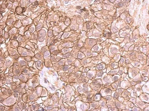
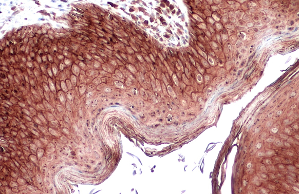
E-cadherin antibody detects E-cadherin protein at membrane on human breast cancer by immunohistochemical analysis.
Sample: Paraffin-embedded breast cancer.
E-cadherin antibody (GTX100443) dilution: 1:500.
Antigen Retrieval: Trilogy™ (EDTA based, pH 8.0) buffer, 15min
E-Cadherin antibody detects E-Cadherin protein at cell membrane in rat prostate by immunohistochemical analysis.
Sample: Paraffin-embedded rat prostate.
E-Cadherin antibody (GTX100443) diluted at 1:500.
Antigen Retrieval: Citrate buffer, pH 6.0, 15 min

Immunohistochemical analysis of paraffin-embedded human ulcerative colitis tissue using E-Cadherin antibody (GTX100443).
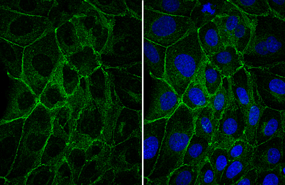
E-Cadherin antibody detects E-Cadherin protein at cell membrane by immunofluorescent analysis.Sample: HCT116 cells were fixed in 4% paraformaldehyde at RT for 15 min.Green: E-Cadherin stained by E-Cadherin antibody (GTX100443) diluted at 1:500.Blue: Fluoroshield with DAPI (GTX30920).Scale bar= 10 μm.
E-cadherin antibody immunoprecipitates E-cadherin protein in IP experiments.
IP samples: MCF-7 whole cell extract
A. Control with 3 μg of preimmune Rabbit IgG
B. Immunoprecipitation of E-cadherin protein by 3 μg E-cadherin antibody (GTX100443)
5 % SDS-PAGE
The immunoprecipitated E-cadherin protein was detected by E-cadherin antibody (GTX100443) diluted at 1:500.
[EasyBlot anti-rabbit IgG (GTX221666-01) was used as a secondary reagent]
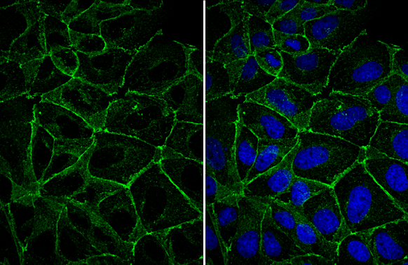
E-Cadherin antibody detects E-Cadherin protein at cell membrane by immunofluorescent analysis.Sample: MDCK cells were fixed in 4% paraformaldehyde at RT for 15 min.Green: E-Cadherin stained by E-Cadherin antibody (GTX100443) diluted at 1:500.
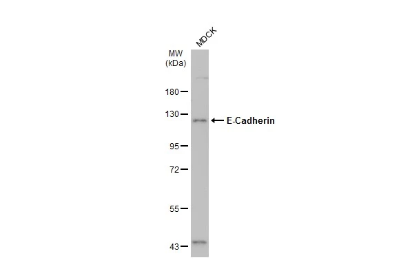
Whole cell extracts (30 μg) was separated by 7.5% SDS-PAGE, and the membrane was blotted with E-Cadherin antibody (GTX100443) diluted at 1:10000. The HRP-conjugated anti-rabbit IgG antibody (GTX213110-01) was used to detect the primary antibody.
E-Cadherin antibody detects E-Cadherin protein at cell membrane by immunofluorescent analysis.Sample: MDCK cells were fixed in 4% paraformaldehyde at RT for 15 min.Green: E-Cadherin stained by E-Cadherin antibody (GTX100443) diluted at 1:500.
E-Cadherin antibody detects E-Cadherin protein at cell membrane and cytoplasm by immunohistochemical analysis.Sample: Paraffin-embedded dog skin.E-Cadherin stained by E-Cadherin antibody (GTX100443) diluted at 1:500.Antigen Retrieval: Citrate buffer, pH 6.0, 15 min
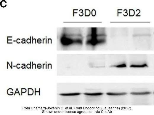
The data was published in the journal Front Endocrinol (Lausanne) in 2017. PMID: 29109696
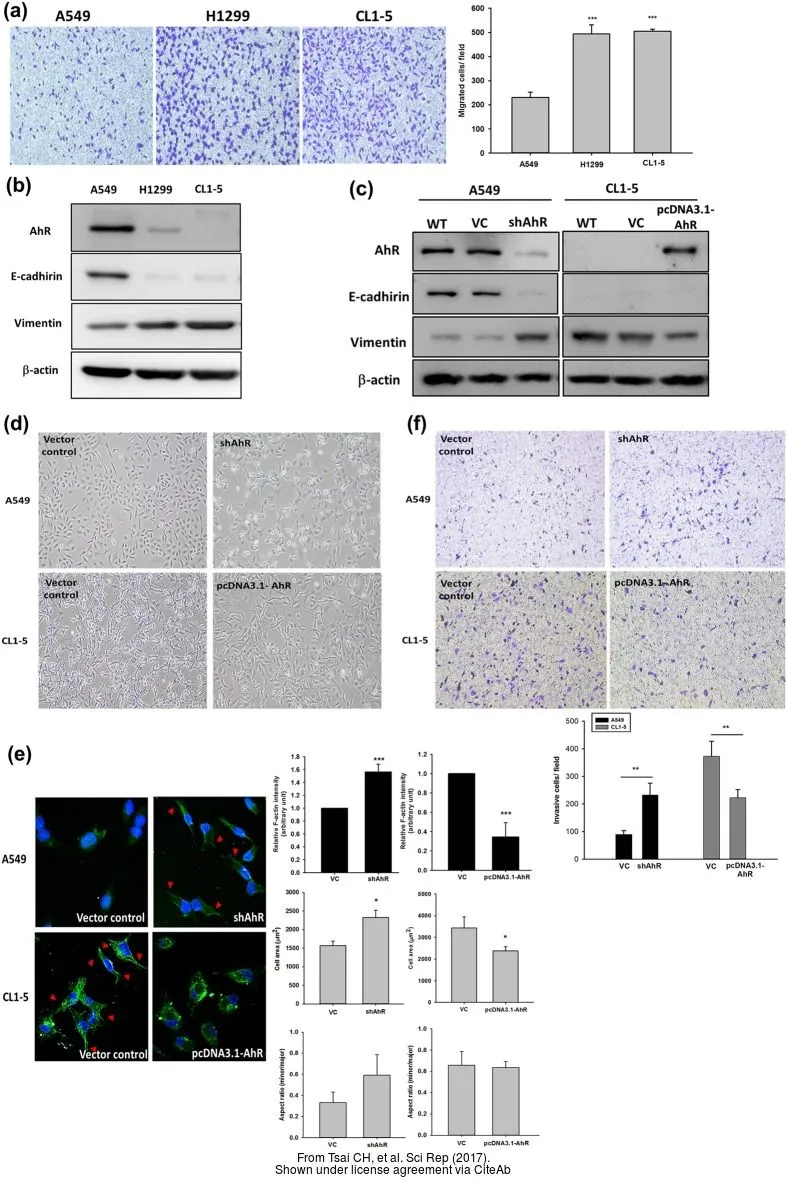
The data was published in the journal Sci Rep in 2017. PMID: 28195146
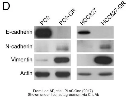
The data was published in the journal PLoS One in 2017. PMID: 28683123
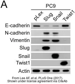
The data was published in the journal PLoS One in 2017. PMID: 28683123
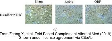
The data was published in the journal Evid Based Complement Alternat Med in 2019. PMID: 31186661
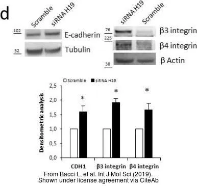
The data was published in the journal Int J Mol Sci in 2019.PMID: 31426484
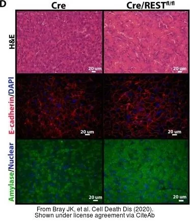
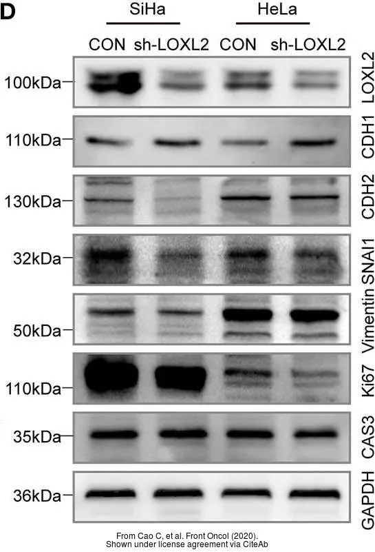
The data was published in the journal Cell Death Dis in 2020.PMID: 32080178
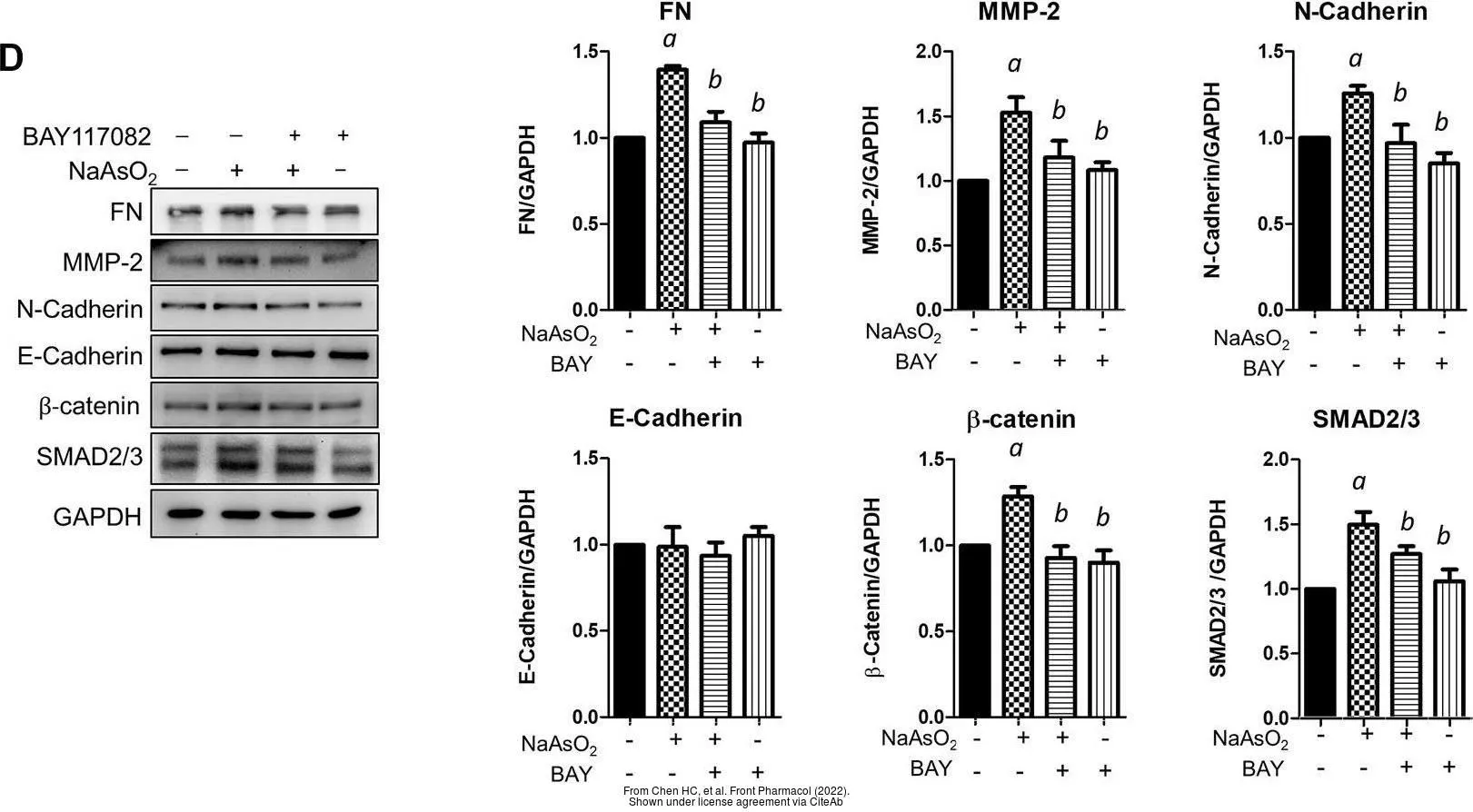
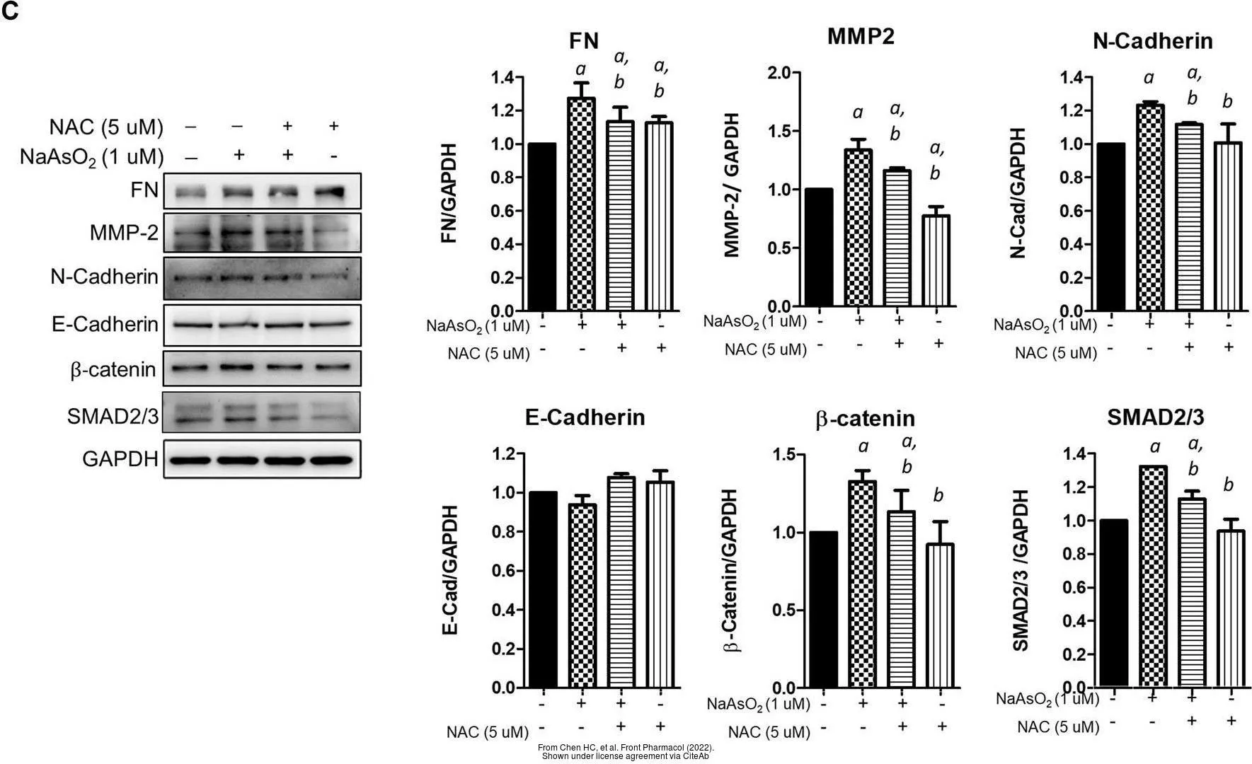
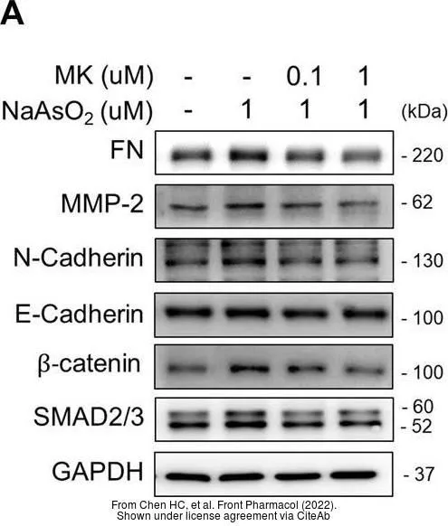
The data was published in the 2022 in Front Pharmacol. PMID: 35517780
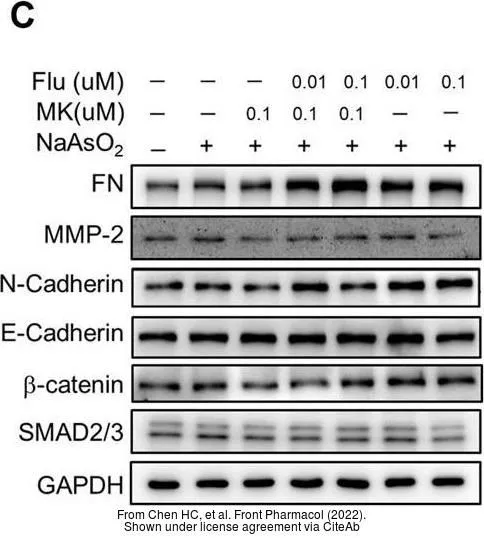
The data was published in the 2022 in Front Pharmacol. PMID: 35517780
The data was published in the 2022 in Front Pharmacol. PMID: 35517780
-
HostRabbit
-
ClonalityPolyclonal
-
IsotypeIgG
-
ApplicationsWB ICC/IF IHC-P IHC-Wm IP PLA
-
ReactivityHuman, Mouse, Rat, Zebrafish, Dog