EAAT1 antibody
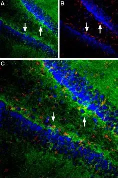
IHC-Frfl (free floating) analysis of perfusion-fixed rat hippocampus tissue using GTX18175 EAAT1 antibody. DAPI is used as a general cell marker (blue).
Panel A : EAAT1 staining (green) is particularly intense along the subgranular layer (arrows)
Panel B : Staining with mouse anti glial fibrillary acidic protein (red)
Panel C : Merged picture confirms presence of densely packed astrocytes along the subgranular layer glial fibrillary acidic protein
Dilution : 1:100
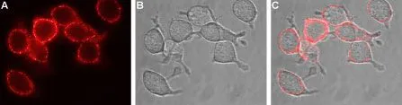
Live cell imaging analysis of live intact U-87 MG cells using GTX18175 EAAT1 antibody.
Panel A : Primary antibody (Red)
Panel B : Live view of the cell
Panel C : Merged images of Panel A and B
Dilution : 1:100
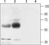
WB analysis of rat (lanes 1 and 3) and mouse (lanes 2 and 4) brain membrane lysates using GTX18175 EAAT1 antibody.
Lane 1, 2 : Primary antibody
Lane 3, 4 : Primary antibody preincubated with immunogen peptide
Dilution : 1:500
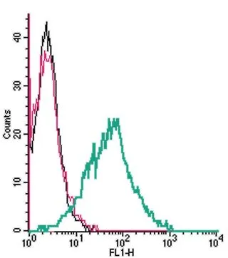
FACS analysis of live intact MEG-01 cells using GTX18175 EAAT1 antibody.
Black : Unstained cell
Red : Cell staining with isotype control antibody
Green : Cell staining with primary antibody
Antibody amount : 2.5 μg
-
HostRabbit
-
ClonalityPolyclonal
-
IsotypeIgG
-
ApplicationsWB ICC/IF FCM IHC (Free Floating) LCI
-
ReactivityHuman, Mouse, Rat