APPLICATION
Application Note
*Optimal dilutions/concentrations should be determined by the researcher.
Application |
Recommended Dilution |
2-4 μg/ml |
2 μg/ml |
Assay dependent |
3-5 μg/10⁶ cells |
Assay dependent |
Assay dependent |
Not tested in other applications.
Calculated MW
Product Note
This antibody shows no cross-reactivity to S1P3. Immunofluorescence data demonstrates that S1P1 is localized to the plasma membrane.
PROPERTIES
Form
Liquid
Buffer
PBS, 0.1% BSA
Preservative
0.05% Sodium azide
Storage
Store as concentrated solution. Centrifuge briefly prior to opening vial. For short-term storage (1-2 weeks), store at 4ºC. For long-term storage, aliquot and store at -20ºC or below. Avoid multiple freeze-thaw cycles.
Concentration
1 mg/ml (Please refer to the vial label for the specific concentration.)
Antigen Species
Mouse
Immunogen
Synthetic peptide corresponding to residues S(359) H P Q K D D G D N P E T I(372) of mouse S1P1.
Purification
Purified by antigen-affinity chromatography
Conjugation
Unconjugated
Note
For laboratory research use only. Not for any clinical, therapeutic, or diagnostic use in humans or animals. Not for animal or human consumption.
Purchasers shall not, and agree not to enable third parties to, analyze, copy, reverse engineer or otherwise attempt to determine the structure or sequence of the product.
TARGET
Synonyms
sphingosine-1-phosphate receptor 1 , CD363 , CHEDG1 , D1S3362 , ECGF1 , EDG-1 , EDG1 , S1P1
Cellular Localization
Cell membrane; Multi-pass membrane protein,Endosome
Background
The protein encoded by this gene is structurally similar to G protein-coupled receptors and is highly expressed in endothelial cells. It binds the ligand sphingosine-1-phosphate with high affinity and high specificity, and suggested to be involved in the processes that regulate the differentiation of endothelial cells. Activation of this receptor induces cell-cell adhesion. Alternative splicing results in multiple transcript variants. [provided by RefSeq, Mar 2016]
Database
Research Area
DATA IMAGES
|
GTX11424 ICC/IF Image
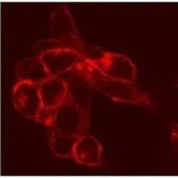
ICC/IF analysis of S1P1 transfected CCL39 cells using GTX11424 EDG1 antibody.
|
|
GTX11424 ICC/IF Image
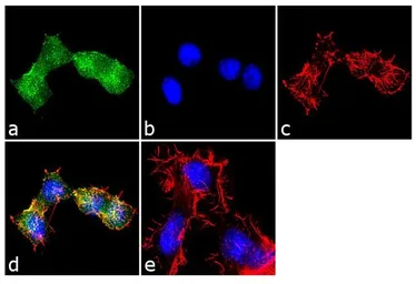
ICC/IF analysis of Neuro-2a cells using GTX11424 EDG1 antibody. Panel e is a no primary antibody control. Green : Primary antibody Blue : Nuclei Red : Actin Fixation : 4% paraformaldehyde Permeabilization : 0.1% Trito X-100 for 10 minutes Dilution : 2 μg/ml in 0.1% BSA and incubated for 3 hours at room temperature
|
|
GTX11424 WB Image
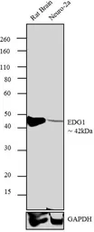
WB analysis of rat Brain (Lane 1) and Neuro- 2a (Lane 2) lysate using GTX11424 EDG1 antibody. Dilution : 2 μg/ml
|
|
GTX11424 WB Image
WB analysis of transfected HEK293 cell lysate using GTX11424 EDG1 antibody.
|
REFERENCE
There are currently no references for EDG1 antibody (GTX11424). Be the first to share your publications with this product.
REVIEW
There are currently no reviews for EDG1 antibody (GTX11424). Be the first to share your experience with this product.