EGFR (phospho Tyr1086) antibody

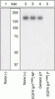
WB (peptide competition) analysis of A431 cells stimulated with 200 ng/mL EGF for 15 minutes (Lane 2-5) using GTX25650 EGFR (phospho Tyr1086) antibody prior incubated with the non-phosphopeptide corresponding to the phosphopeptide immunogen (Lane 3), a generic phosphotyrosine-containing peptide (Lane 4), or the phosphopeptide immunogen (Lane 5). The data show that only the immunogen phosphopeptide blocks the signal, demonstrating the specificity of the antibody.
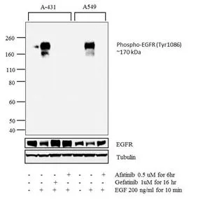
WB analysis of membrane enriched extracts (30 ug lysate) of A-431 (Lane 1) treated with EGF (200ng/ml for 10 minutes) (Lane 2), A-431 treated with Gefitinib followed by EGF (1uM for 16 hours, 200ng/ml for 10 minutes) (Lane 3), A-431 treated with Afatinib followed by EGF (0.5 uM for 6 hours, 200ng/ml for 10 minutes) (Lane 4), A549 (Lane 5) treated with EGF (200ng/ml for 10 minutes) (Lane 6), and A549 treated with Afatinib followed by EGF (0.5 uM for 6 hours, 200ng/ml for 10 minutes) (Lane 7) using GTX25650 EGFR (phospho Tyr1086) antibody.
Dilution : 1:1000
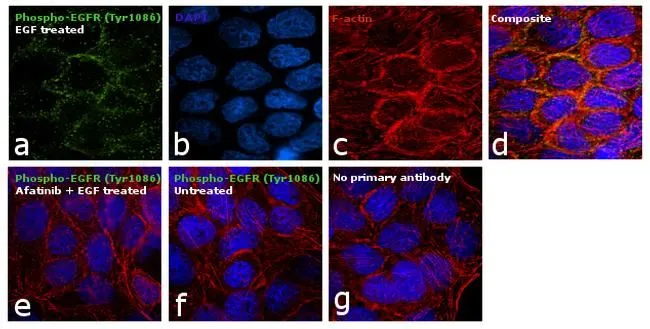
ICC/IF analysis of A431 cells treated with Afatinib (1uM for 6hrs) followed by EGF (200ng/ml for 10 minutes) using GTX25650 EGFR (phospho Tyr1086) antibody.
Green : Primary antibody
Blue : Nuclei
Red : Actin
Fixation : 4% paraformaldehyde
Permeabilization : 0.1% Triton X-100 for 15 minutes
Dilution : 1:100 in 0.1% BSA incubated at 4ºC
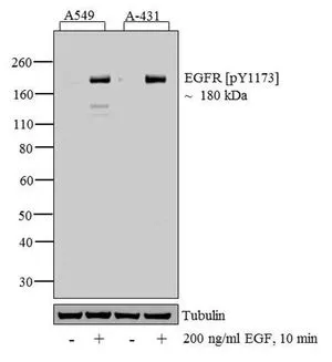
WB analysis of 30 μg of A549 (Lane 1) treated for 10 minutes with 200 ng/ml of EGF (Lane 2), A431 (Lane 3) treated for 10 minutes with 200 ng/ml of EGF (Lane 4) cell lysate using GTX25650 EGFR (phospho Tyr1086) antibody.
Dilution : 1:1000
-
HostRabbit
-
ClonalityPolyclonal
-
IsotypeIgG
-
ApplicationsWB ICC/IF IHC
-
ReactivityHuman, Mouse