EIF2 beta antibody

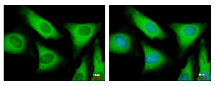
EIF2 beta antibody detects EIF2S2 protein at cytoplasm by immunofluorescent analysis.
Sample: HeLa cells were fixed in 4% paraformaldehyde at RT for 15 min.
Green: EIF2S2 protein stained by EIF2 beta antibody (GTX106484) diluted at 1:500.
Blue: Hoechst 33343 staining.
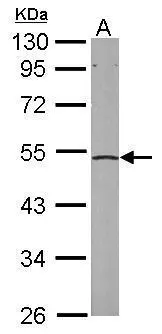
Sample (50 ug of whole cell lysate)
A: mouse liver
10% SDS PAGE
GTX106484 diluted at 1:1000
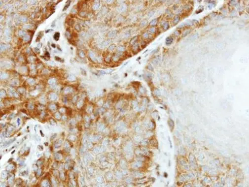
Immunohistochemical analysis of paraffin-embedded SG xenograft, using EIF2 beta(GTX106484) antibody at 1:500 dilution.
Antigen Retrieval: Citrate buffer, pH 6.0, 15 min
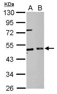
EIF2 beta antibody detects EIF2S2 protein by Western blot analysis.
A. 30 μg PC-12 whole cell lysate/extract
B. 30 ug Rat2 whole cell lysate/extract
10 % SDS-PAGE
EIF2 beta antibody (GTX106484) dilution: 1:2000
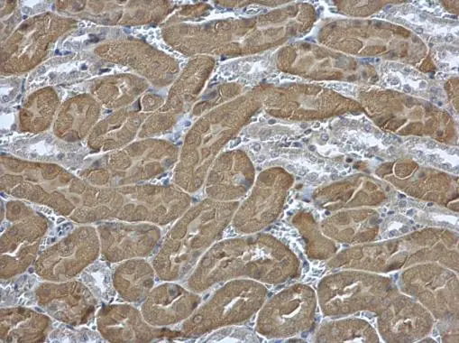
EIF2 beta antibody detects EIF2 beta protein at cytoplasm in mouse kidney by immunohistochemical analysis.
Sample: Paraffin-embedded mouse kidney.
EIF2 beta antibody (GTX106484) diluted at 1:500.
Antigen Retrieval: Citrate buffer, pH 6.0, 15 min
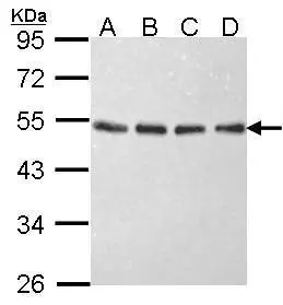
Sample (30 ug of whole cell lysate)
A: 293T
B: A431 (GTX27909)
C: H1299
D: Hela
10% SDS PAGE
GTX106484 diluted at 1:1000
-
HostRabbit
-
ClonalityPolyclonal
-
IsotypeIgG
-
ApplicationsWB ICC/IF IHC-P IP
-
ReactivityHuman, Mouse, Rat