ERK2 antibody

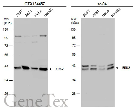
Various whole cell extracts (30 μg) were separated by 10% SDS-PAGE, and the membrane was blotted with ERK2 antibody (GTX134457) diluted at 1:1000 and competitor's antibody (sc-94) diluted at 1:2000. The HRP-conjugated anti-rabbit IgG antibody (GTX213110-01) was used to detect the primary antibody.
*The competitor is not affiliated with GeneTex and does not endorse this product.
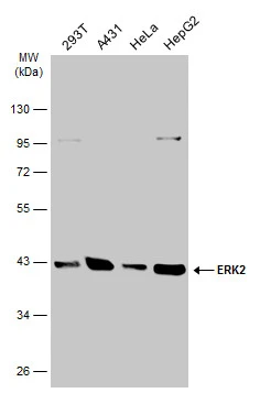
Various whole cell extracts (30 μg) were separated by 10% SDS-PAGE, and the membrane was blotted with ERK2 antibody (GTX134457) diluted at 1:1000. The HRP-conjugated anti-rabbit IgG antibody (GTX213110-01) was used to detect the primary antibody.
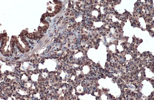
ERK2 antibody detects ERK2 protein at cytoplasm and nucleus by immunohistochemical analysis.Sample: Paraffin-embedded mouse lung.ERK2 stained by ERK2 antibody (GTX134457) diluted at 1:500.Antigen Retrieval: Citrate buffer, pH 6.0, 15 min
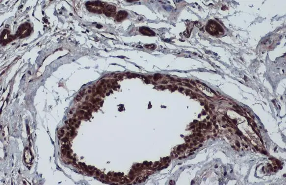
ERK2 antibody detects ERK2 protein at cytoplasm and nucleus by immunohistochemical analysis.Sample: Paraffin-embedded cat mammary gland.ERK2 stained by ERK2 antibody (GTX134457) diluted at 1:500.Antigen Retrieval: Citrate buffer, pH 6.0, 15 min
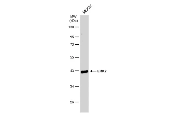
Whole cell extract (30 μg) was separated by 10% SDS-PAGE, and the membrane was blotted with ERK2 antibody (GTX134457) diluted at 1:1000. The HRP-conjugated anti-rabbit IgG antibody (GTX213110-01) was used to detect the primary antibody, and the signal was developed with Trident ECL plus-Enhanced.
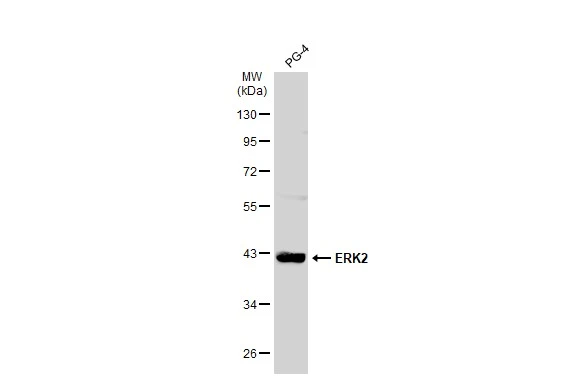
Whole cell extract (30 μg) was separated by 10% SDS-PAGE, and the membrane was blotted with ERK2 antibody (GTX134457) diluted at 1:1000. The HRP-conjugated anti-rabbit IgG antibody (GTX213110-01) was used to detect the primary antibody.
-
HostRabbit
-
ClonalityPolyclonal
-
IsotypeIgG
-
ApplicationsWB IHC-P
-
ReactivityHuman, Mouse, Cat, Dog