EXOSC8 antibody
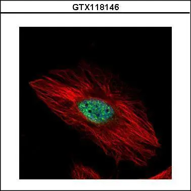
Confocal immunofluorescence analysis (Olympus FV10i) of methanol-fixed HeLa, using EXOSC8(GTX118146) antibody (Green) at 1:500 dilution. Alpha-tubulin filaments were labeled with GTX11304 (Red) at 1:2000.
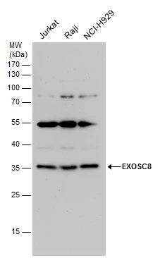
EXOSC8 antibody detects EXOSC8 protein by western blot analysis. Various whole cell extracts (30 μg) were separated by 12% SDS-PAGE, and the membrane was blotted with EXOSC8 antibody (GTX118146) diluted by 1:1000.
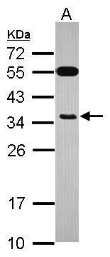
Sample (30 ug of whole cell lysate)
A: HepG2
12% SDS PAGE
GTX118146 diluted at 1:1000
-
HostRabbit
-
ClonalityPolyclonal
-
IsotypeIgG
-
ApplicationsWB ICC/IF
-
ReactivityHuman