FEN1 antibody
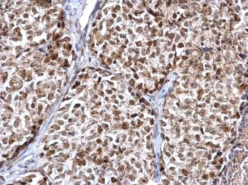
FEN1 antibody detects FEN1 protein at nucleus on human breast carcinoma by immunohistochemical analysis.
Sample: Paraffin-embedded human breast carcinoma.
FEN1 antibody (GTX101777) diluted at 1:500.
Antigen Retrieval: Trilogy™ (EDTA based, pH 8.0) buffer, 15min
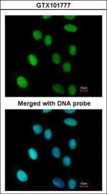
Immunofluorescence analysis of paraformaldehyde-fixed A549, using FEN1(GTX101777) antibody at 1:200 dilution.
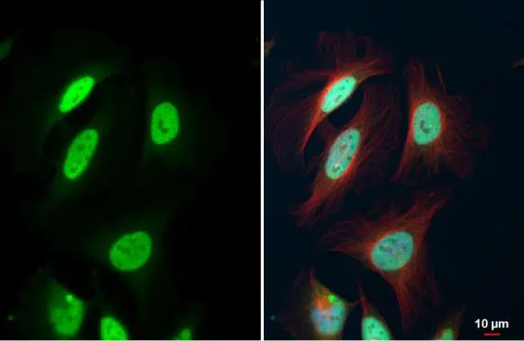
FEN1 antibody detects FEN1 protein at nucleus by immunofluorescent analysis.Sample: HeLa cells were fixed in 4% paraformaldehyde at RT for 15 min.Green: FEN1 stained by FEN1 antibody (GTX101777) diluted at 1:500.Red: alpha Tubulin, a cytoskeleton marker, stained by alpha Tubulin antibody [GT114] (GTX628802) diluted at 1:1000.Blue: Fluoroshield with DAPI (GTX30920).
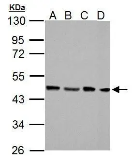
FEN1 antibody detects FEN1 protein by western blot analysis.
A. 30 μg NIH-3T3 whole cell lysate/extract
B. 30 μg JC whole cell lysate/extract
C. 30 μg BCL-1 whole cell lysate/extract
D. 30 μg Raw264.7 whole cell lysate/extract
10% SDS-PAGE
FEN1 antibody (GTX101777) dilution: 1:1000
The HRP-conjugated anti-rabbit IgG antibody (GTX213110-01) was used to detect the primary antibody.
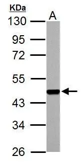
FEN1 antibody detects FEN1 protein by western blot analysis.
A. 30 μg PC-12 whole cell lysate/extract
10% SDS-PAGE
FEN1 antibody (GTX101777) dilution: 1:1000
The HRP-conjugated anti-rabbit IgG antibody (GTX213110-01) was used to detect the primary antibody.
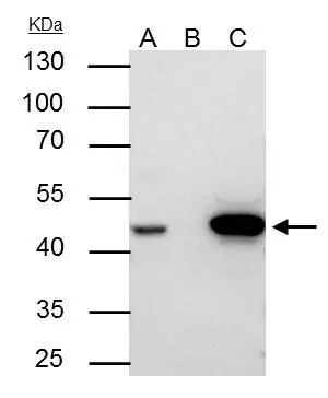
FEN1 antibody immunoprecipitates FEN1 protein in IP experiments.
IP samples: Jurkat whole cell extract
A. 30 μg Jurkat whole cell extract
B. Control with 4 μg of preimmune Rabbit IgG
C. Immunoprecipitation of FEN1 protein by 4 μg FEN1 antibody (GTX101777)
10 % SDS-PAGE
The immunoprecipitated FEN1 protein was detected by FEN1 antibody (GTX101777) diluted at 1:500.
[EasyBlot anti-rabbit IgG (GTX221666-01) was used as a secondary reagent]
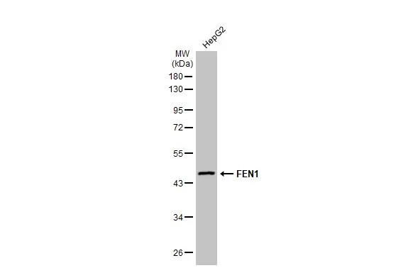
Whole cell extract (30 μg) was separated by 10% SDS-PAGE, and the membrane was blotted with FEN1 antibody (GTX101777) diluted at 1:1000. The HRP-conjugated anti-rabbit IgG antibody (GTX213110-01) was used to detect the primary antibody, and the signal was developed with Trident ECL plus-Enhanced.
-
HostRabbit
-
ClonalityPolyclonal
-
IsotypeIgG
-
ApplicationsWB ICC/IF IHC-P IP IHC PLA
-
ReactivityHuman, Mouse, Rat