Fascin 1 antibody

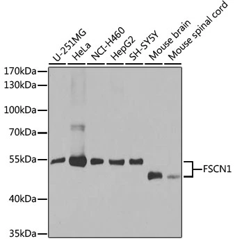
WB analysis of various sample lysates using GTX55616 Fascin 1 antibody.
Dilution : 1:1000
Loading : 25μg per lane
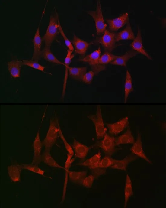
ICC/IF analysis of NIH/3T3 cells using GTX55616 Fascin 1 antibody.
Blue : DAPI
Dilution : 1:100
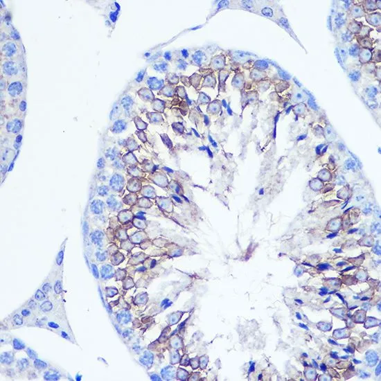
IHC-P analysis of mouse testis tissue using GTX55616 Fascin 1 antibody.
Dilution : 1:100
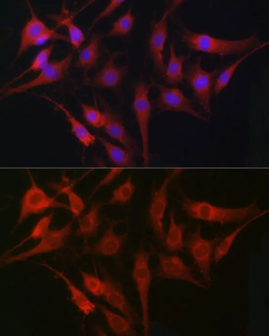
ICC/IF analysis of C6 cells using GTX55616 Fascin 1 antibody.
Blue : DAPI
Dilution : 1:100
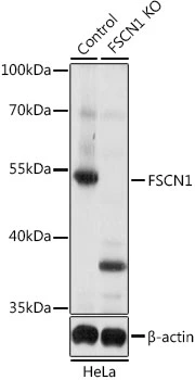
WB analysis of normal (control) and knockout (KO) HeLa cell lysate using GTX55616 Fascin 1 antibody.
Dilution : 1:1000
Loading : 25μg per lane
-
HostRabbit
-
ClonalityPolyclonal
-
IsotypeIgG
-
ApplicationsWB ICC/IF IHC-P
-
ReactivityHuman, Mouse, Rat