Fibrillarin antibody

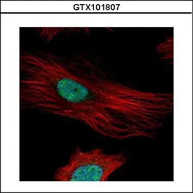
Confocal immunofluorescence analysis (Olympus FV10i) of paraformaldehyde-fixed HeLa, using Fibrillarin(GTX101807) antibody (Green) at 1:500 dilution. Alpha-tubulin filaments were labeled with GTX11304 (Red) at 1:2000.
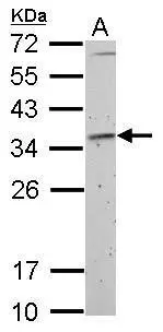
Fibrillarin antibody detects FBL protein by Western blot analysis.
A. 30 μg Rat2 whole cell lysate/extract
12 % SDS-PAGE
Fibrillarin antibody (GTX101807) dilution: 1:1000
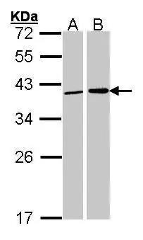
Sample (30 ug of whole cell lysate)
A: 293T
B: H1299
12% SDS PAGE
GTX101807 diluted at 1:1000
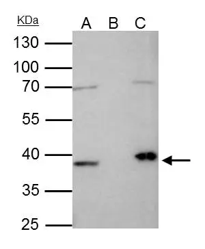
Fibrillarin antibody immunoprecipitates FBL protein in IP experiments.
IP samples: HeLa nuclear extract
A. 35 μg HeLa nuclear extract
B. Control with 4 μg of preimmune Rabbit IgG
C. Immunoprecipitation of FBL protein by 4 μg Fibrillarin antibody (GTX101807)
10 % SDS-PAGE
The immunoprecipitated FBL protein was detected by Fibrillarin antibody (GTX101807) diluted at 1:1000.
[EasyBlot anti-rabbit IgG (GTX221666-01) was used as a secondary reagent]

Sample (30 ug of whole cell lysate)
A:NIH-3T3 10% SDS PAGE
GTX101807 diluted at 1:1000
-
HostRabbit
-
ClonalityPolyclonal
-
IsotypeIgG
-
ApplicationsWB ICC/IF IP
-
ReactivityHuman, Mouse, Rat